Application
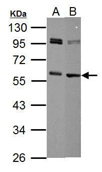
Sample (30 µg of whole cell lysate) A: NIH-3T3 B: Raw264.7 10% SDS PAGE diluted at 1:1000 The HRP-conjugated anti-rabbit IgG antibody was used to detect the primary antibody.Application
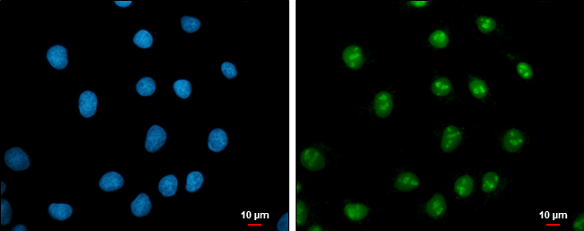
c-Myc antibody detects c-Myc protein at nucleus by immunofluorescent analysis. Sample: NT2D1 cells were fixed in 4% paraformaldehyde at RT for 15 min. Green: c-Myc protein stained by c-Myc antibody diluted at 1:200. Blue: Hoechst 33342 staining. Scale bar = 10 μm.Application
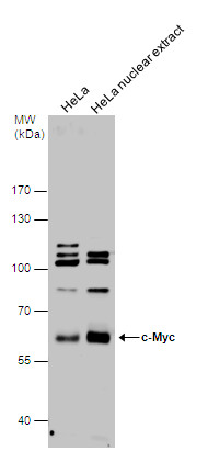
c-Myc antibody detects c-Myc protein by western blot analysis. HeLa whole cell extracts and nuclear extracts (30 µg) were separated by 7.5% SDS-PAGE, and the membrane was blotted with c-Myc antibody diluted at 1:1000. The HRP-conjugated anti-rabbit IgG antibody was used to detect the primary antibody.Application
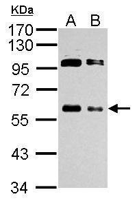
Sample (whole cell lysate) A: 293T 20ug B: 293T 10ug 10% SDS PAGE diluted at 1:1000 The HRP-conjugated anti-rabbit IgG antibody was used to detect the primary antibody.Application
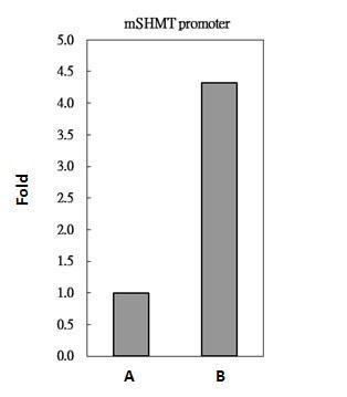
c-Myc antibody immunoprecipitates c-Myc protein-DNA in ChIP experiments. ChIP Sample: 293T whole cell lysate/extract A. 5 μg preimmune rabbit IgG B. 5 μg of c-Myc antibody The precipitated DNA was detected by PCR with primer set targeting to mSHMT promoter.Application
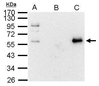
c-Myc antibody immunoprecipitates c-Myc protein in IP experiments. IP Sample: 1000 μg 293T whole cell lysate/extract A. 20 μg 293T whole cell lysate/extract B. Control with 2.5 μg of preimmune rabbit IgG C. Immunoprecipitation of c-Myc protein by 2.5 μg of c-Myc antibody 10% SDS-PAGE The immunoprecipitated c-Myc protein was detected by c-Myc antibody diluted at 1:1000. EasyBlot anti-rabbit IgG was used as a secondary reagent.| Product Name | c-Myc antibody |
|---|---|
| Antibody Type | Tags Antibodies |
| Product description | Rabbit Polyclonal antibody to c-Myc (v-myc myelocytomatosis viral oncogene homolog (avian))The protein encoded by this gene is a multifunctional, nuclear phosphoprotein that plays a role in cell cycle progression, apoptosis and cellular transformation. It functions as a transcription factor that regulates transcription of specific target genes. Mutations, overexpression, rearrangement and translocation of this gene have been associated with a variety of hematopoietic tumors, leukemias and lymphomas, including Burkitt lymphoma. There is evidence to show that alternative translation initiations from an upstream, in-frame non-AUG (CUG) and a downstream AUG start site result in the production of two isoforms with distinct N-termini. The synthesis of non-AUG initiated protein is suppressed in Burkitt's lymphomas, suggesting its importance in the normal function of this gene. [provided by RefSeq] |
| Immunogen | Recombinant protein encompassing a sequence within the center region of human c-Myc. The exact sequence is proprietary. |
| Clonality | Polyclonal |
|---|---|
| Isotype | IgG |
| Host Species | Rabbit |
| Tested Applications | ICC/IFIPWBChIP |
| *Optimal dilutions/concentrations should be determined by the researcher.
: |
|
| Species Reactivity | HumanMouse |
| Predicted Reactivity | Rat, Dog, Cat, Pig, Rabbit, Sheep, Rhesus Monkey, Chimpanzee, BovineRat (86%), Dog (93%), Cat (92%), Pig (94%), Rabbit (93%), Sheep (92%), Rhesus Monkey (98%), Chimpanzee (99%), Bovine (92%) |
| Concentration | 1mg/ml (Please refer to the vial label for the specific concentration) |
| Purification | Affinity purified |
| Gene Synonyms | MRTL antibody bHLHe39 antibody c-Myc antibody MYC antibody avian myelocytomatosis viral oncogene homolog antibody myc-related translation/localization regulatory factor antibody class E basic helix-loop-helix protein 39 antibody transcription factor |
|---|---|
| Molecular Weight(MW) | 49 kDa (note) |
| Cellular Localization | Nucleus |
Application
Sample (30 µg of whole cell lysate) A: NIH-3T3 B: Raw264.7 10% SDS PAGE diluted at 1:1000 The HRP-conjugated anti-rabbit IgG antibody was used to detect the primary antibody.
Application
c-Myc antibody detects c-Myc protein at nucleus by immunofluorescent analysis. Sample: NT2D1 cells were fixed in 4% paraformaldehyde at RT for 15 min. Green: c-Myc protein stained by c-Myc antibody diluted at 1:200. Blue: Hoechst 33342 staining. Scale bar = 10 μm.
Application
c-Myc antibody detects c-Myc protein by western blot analysis. HeLa whole cell extracts and nuclear extracts (30 µg) were separated by 7.5% SDS-PAGE, and the membrane was blotted with c-Myc antibody diluted at 1:1000. The HRP-conjugated anti-rabbit IgG antibody was used to detect the primary antibody.
Application
Sample (whole cell lysate) A: 293T 20ug B: 293T 10ug 10% SDS PAGE diluted at 1:1000 The HRP-conjugated anti-rabbit IgG antibody was used to detect the primary antibody.
Application
c-Myc antibody immunoprecipitates c-Myc protein-DNA in ChIP experiments. ChIP Sample: 293T whole cell lysate/extract A. 5 μg preimmune rabbit IgG B. 5 μg of c-Myc antibody The precipitated DNA was detected by PCR with primer set targeting to mSHMT promoter.
Application
c-Myc antibody immunoprecipitates c-Myc protein in IP experiments. IP Sample: 1000 μg 293T whole cell lysate/extract A. 20 μg 293T whole cell lysate/extract B. Control with 2.5 μg of preimmune rabbit IgG C. Immunoprecipitation of c-Myc protein by 2.5 μg of c-Myc antibody 10% SDS-PAGE The immunoprecipitated c-Myc protein was detected by c-Myc antibody diluted at 1:1000. EasyBlot anti-rabbit IgG was used as a secondary reagent.| Positive Control | 293T , NIH-3T3 , Raw264.7 , HeLa , HeLa nuclear extract |
|---|---|
| Application Notes | *Optimal dilutions/concentrations should be determined by the researcher.
: |
| Form | Liquid |
|---|---|
| Storage Instructions | Keep as concentrated solution. Aliquot and store at -20ºC or below. Avoid multiple freeze-thaw cycles. |
| Storage Buffer | 1XPBS, 20% Glycerol (pH7). 0.01% Thimerosal was added as a preservative. |


