Application
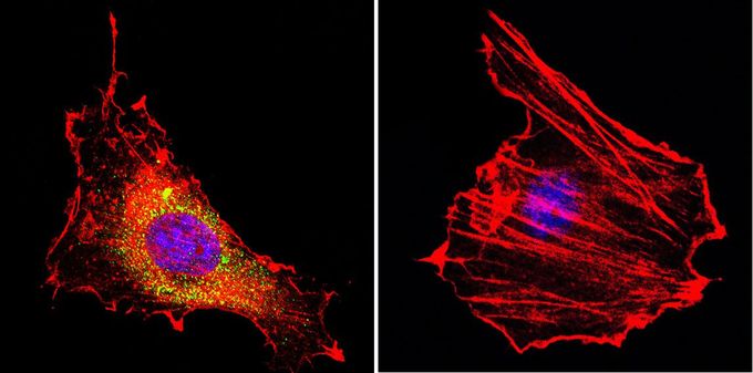
Immunofluorescent analysis of CNG Channel in B-3 cells. CNG Channel staining (green), F-Actin staining with Phalloidin (red) and nuclei with DAPI (blue) is shown. Cells were grown on slides and fixed with formaldehyde prior to staining. Cells were probed without (control) or with CNG Channel antibody [CNC 9C1] at a dilution of 1:20 over night at 4 ?C, washed with PBS and incubated with a proper secondary antibody. Images were taken at 60X magnification.| Product Name | CNG Channel antibody |
|---|---|
| Antibody Type | Primary Antibodies |
| Product description | Mouse monoclonal antibody [CNC 9C1] to CNG ChannelThe protein encoded by this gene is involved in phototransduction. Along with another protein, the encoded protein forms a cGMP-gated cation channel in the plasma membrane, allowing depolarization of rod photoreceptors. This represents the last step in the phototransduction pathway. Defects in this gene are a cause of retinitis pigmentosa autosomal recessive (ARRP) disease. Two transcript variants encoding different isoforms have been found for this gene. [provided by RefSeq, Dec 2008] |
| Immunogen | MBP-fusion protein of the C-terminal region of the a2 subunit (amino acids K581-D670) |
| Clonality | Monoclonal |
|---|---|
| Isotype | IgG1 |
| Clone Number | CNC 9C1 |
| Host Species | Mouse |
| Tested Applications | ICC/IFIHCWB |
| ICC/IF: 1:10 - 1:100: |
|
| Species Reactivity | HumanMouseRat |
| Concentration | 1mg/ml (Please refer to the vial label for the specific concentration) |
| Gene Synonyms | cyclic nucleotide gated channel alpha 1 CNCG1 CNCG RCNCalpha RCNCa CNG-1 RCNC1 RP49 CNGA1 CNG1 |
|---|---|
| Target | CNG Channel |
| Tissue Specificity | This antibody is specific to the a2 subunit of the CNG channel. |
Application
Immunofluorescent analysis of CNG Channel in B-3 cells. CNG Channel staining (green), F-Actin staining with Phalloidin (red) and nuclei with DAPI (blue) is shown. Cells were grown on slides and fixed with formaldehyde prior to staining. Cells were probed without (control) or with CNG Channel antibody [CNC 9C1] at a dilution of 1:20 over night at 4 ?C, washed with PBS and incubated with a proper secondary antibody. Images were taken at 60X magnification.| Application Notes | ICC/IF: 1:10 - 1:100: |
|---|
| Form | Liquid |
|---|---|
| Storage Instructions | -20° C, Avoid Freeze/Thaw Cycles |
| Storage Buffer | Ascites containing 0.05 % sodium azide |


