Application
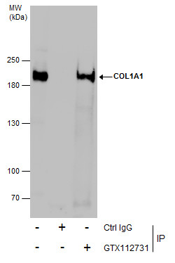
Immunoprecipitation of COL1A1 protein from SK-N-AS whole cell extracts using 5 μg of COL1A1 antibody [N1N2], N-term . Western blot analysis was performed using COL1A1 antibody [N1N2], N-term . EasyBlot anti-Rabbit IgG was used as a secondary reagent.Application
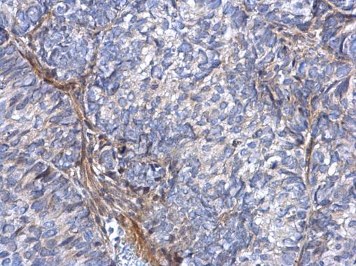
COL1A1 antibody [N1N2], N-term detects COL1A1 protein at secreted on human cervical carcinoma by immunohistochemical analysis. Sample: Paraffin-embedded human cervical carcinoma. COL1A1 antibody [N1N2], N-term dilution: 1:500.Application
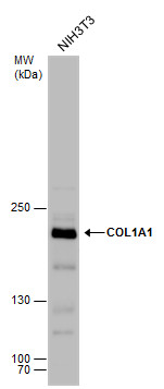
COL1A1 antibody detects COL1A1 protein by western blot analysis. Whole cell extracts (30 µg) was separated by 5% SDS-PAGE, and the membrane was blotted with COL1A1 antibody diluted by 1:500. The HRP-conjugated anti-rabbit IgG antibody was used to detect the primary antibody.Application
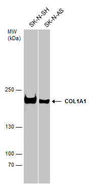
Various whole cell extracts (30 µg) were separated by 5% SDS-PAGE, and the membrane was blotted with COL1A1 antibody [N1N2], N-term diluted at 1:1000. The HRP-conjugated anti-rabbit IgG antibody was used to detect the primary antibody.Application
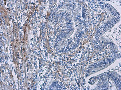
COL1A1 antibody [N1N2], N-term detects COL1A1 protein at secreted in human colon cancer by immunohistochemical analysis. Sample: Paraffin-embedded human colon cancer. COL1A1 antibody [N1N2], N-term diluted at 1:500.Application
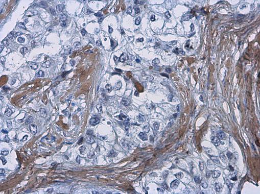
COL1A1 antibody [N1N2], N-term detects COL1A1 protein at secreted in human breast carcinoma by immunohistochemical analysis. Sample: Paraffin-embedded human breast carcinoma. COL1A1 antibody [N1N2], N-term diluted at 1:500.Application
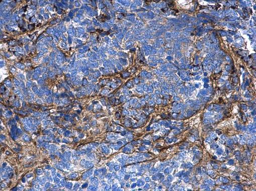
COL1A1 antibody [N1N2], N-term detects COL1A1 protein at secreted on human endometrial carcinoma by immunohistochemical analysis. Sample: Paraffin-embedded human endometrial carcinoma. COL1A1 antibody [N1N2], N-term diluted at 1:500.Application
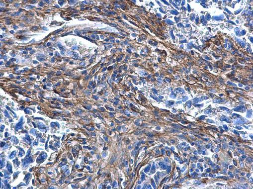
COL1A1 antibody [N1N2], N-term detects COL1A1 protein at secreted on human lung carcinoma by immunohistochemical analysis. Sample: Paraffin-embedded human lung carcinoma. COL1A1 antibody [N1N2], N-term diluted at 1:500.Application
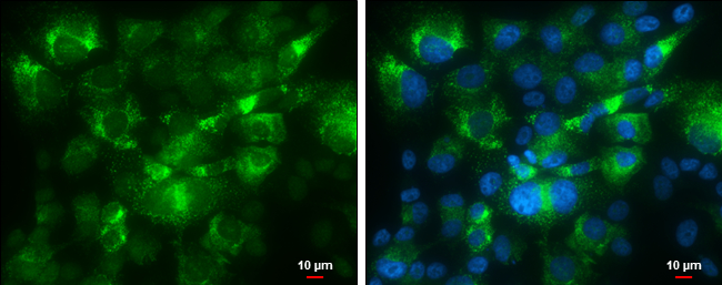
COL1A1 antibody [N1N2], N-term detects COL1A1 protein at cytoplasm by immunofluorescent analysis. Sample: SK-N-AS cells were fixed in 4% paraformaldehyde at RT for 15 min. Green: COL1A1 protein stained by COL1A1 antibody [N1N2], N-term diluted at 1:500. Blue: Hoechst 33342 staining. Scale bar = 10 μm.| Product Name | COL1A1 antibody , N-term |
|---|---|
| Antibody Type | Primary Antibodies |
| Product description | Rabbit Polyclonal antibody to COL1A1 (collagen, type I, alpha 1)This gene encodes the pro-alpha1 chains of type I collagen whose triple helix comprises two alpha1 chains and one alpha2 chain. Type I is a fibril-forming collagen found in most connective tissues and is abundant in bone, cornea, dermis and tendon. Mutations in this gene are associated with osteogenesis imperfecta types I-IV, Ehlers-Danlos syndrome type VIIA, Ehlers-Danlos syndrome Classical type, Caffey Disease and idiopathic osteoporosis. Reciprocal translocations between chromosomes 17 and 22, where this gene and the gene for platelet-derived growth factor beta are located, are associated with a particular type of skin tumor called dermatofibrosarcoma protuberans, resulting from unregulated expression of the growth factor. Two transcripts, resulting from the use of alternate polyadenylation signals, have been identified for this gene. [provided by R. Dalgleish] |
| Immunogen | Recombinant protein encompassing a sequence within the N-terminus region of human COL1A1. The exact sequence is proprietary. |
| Modification | Acetylated |
| Clonality | Polyclonal |
|---|---|
| Isotype | IgG |
| Host Species | Rabbit |
| Tested Applications | ICC/IFIHC-PIPWB |
| WB:1:500~1:1000 IP:1:100~1:500 ICC/IF:1:100~1:500 IHC:1:50~1:500 |
|
| Species Reactivity | HumanMouse |
| Predicted Reactivity | Rat, Xenopus laevis, Dog, BovineRat (94%), Xenopus laevis (83%), Dog (98%), Bovine (98%) |
| Concentration | 1mg/ml (Please refer to the vial label for the specific concentration) |
| Purification | Affinity purified |
| Gene Synonyms | OI4 antibody COL1A1 antibody collagen alpha-1(I) chain antibody pro-alpha-1 collagen type 1 antibody alpha-1 type I collagen antibody |
|---|---|
| Molecular Weight(MW) | 139 kDa (note) |
| Cellular Localization | Secreted , extracellular space , extracellular matrix |
Application
Immunoprecipitation of COL1A1 protein from SK-N-AS whole cell extracts using 5 μg of COL1A1 antibody [N1N2], N-term . Western blot analysis was performed using COL1A1 antibody [N1N2], N-term . EasyBlot anti-Rabbit IgG was used as a secondary reagent.
Application
COL1A1 antibody [N1N2], N-term detects COL1A1 protein at secreted on human cervical carcinoma by immunohistochemical analysis. Sample: Paraffin-embedded human cervical carcinoma. COL1A1 antibody [N1N2], N-term dilution: 1:500.
Application
COL1A1 antibody detects COL1A1 protein by western blot analysis. Whole cell extracts (30 µg) was separated by 5% SDS-PAGE, and the membrane was blotted with COL1A1 antibody diluted by 1:500. The HRP-conjugated anti-rabbit IgG antibody was used to detect the primary antibody.
Application
Various whole cell extracts (30 µg) were separated by 5% SDS-PAGE, and the membrane was blotted with COL1A1 antibody [N1N2], N-term diluted at 1:1000. The HRP-conjugated anti-rabbit IgG antibody was used to detect the primary antibody.
Application
COL1A1 antibody [N1N2], N-term detects COL1A1 protein at secreted in human colon cancer by immunohistochemical analysis. Sample: Paraffin-embedded human colon cancer. COL1A1 antibody [N1N2], N-term diluted at 1:500.
Application
COL1A1 antibody [N1N2], N-term detects COL1A1 protein at secreted in human breast carcinoma by immunohistochemical analysis. Sample: Paraffin-embedded human breast carcinoma. COL1A1 antibody [N1N2], N-term diluted at 1:500.
Application
COL1A1 antibody [N1N2], N-term detects COL1A1 protein at secreted on human endometrial carcinoma by immunohistochemical analysis. Sample: Paraffin-embedded human endometrial carcinoma. COL1A1 antibody [N1N2], N-term diluted at 1:500.
Application
COL1A1 antibody [N1N2], N-term detects COL1A1 protein at secreted on human lung carcinoma by immunohistochemical analysis. Sample: Paraffin-embedded human lung carcinoma. COL1A1 antibody [N1N2], N-term diluted at 1:500.
Application
COL1A1 antibody [N1N2], N-term detects COL1A1 protein at cytoplasm by immunofluorescent analysis. Sample: SK-N-AS cells were fixed in 4% paraformaldehyde at RT for 15 min. Green: COL1A1 protein stained by COL1A1 antibody [N1N2], N-term diluted at 1:500. Blue: Hoechst 33342 staining. Scale bar = 10 μm.| Positive Control | SK-N-SH , SK-N-AS , NIH3T3 |
|---|---|
| Application Notes | WB:1:500~1:1000 IP:1:100~1:500 ICC/IF:1:100~1:500 IHC:1:50~1:500 |
| Form | Liquid |
|---|---|
| Storage Instructions | Keep as concentrated solution. Aliquot and store at -20ºC or below. Avoid multiple freeze-thaw cycles. |
| Storage Buffer | 1XPBS, 1% BSA, 20% Glycerol (pH7). 0.025% ProClin 300 was added as a preservative. |


