Application
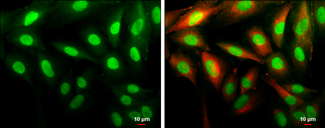
Cyclin D1 antibody [N1C3] detects Cyclin D1 protein at cytoplasm and nucleus by immunofluorescent analysis. Sample: SK-N-SH cells were fixed in 4% paraformaldehyde at RT for 15 min. Green: Cyclin D1 protein stained by Cyclin D1 antibody [N1C3] diluted at 1:500. Red: beta Tubulin 3/ Tuj1, a cytoskeleton marker, stained by beta Tubulin 3/ Tuj1 antibody [GT11710] diluted at 1:500. Scale bar = 10 μm.Application
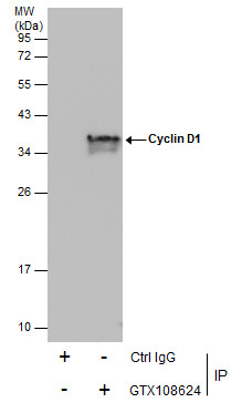
Immunoprecipitation of Cyclin D1 protein from MCF-7 whole cell extracts using 5 μg of Cyclin D1 antibody [N1C3] . Western blot analysis was performed using Cyclin D1 antibody [N1C3] . EasyBlot anti-Rabbit IgG was used as a secondary reagent.Application
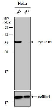
Wild-type (WT) and Cyclin D1 knockout (KO) HeLa cell extracts (30 µg) were separated by 12% SDS-PAGE, and the membrane was blotted with Cyclin D1 antibody [N1C3] diluted at 1:5000. The HRP-conjugated anti-rabbit IgG antibody was used to detect the primary antibody, and the signal was developed with Trident ECL plus-Enhanced.Application
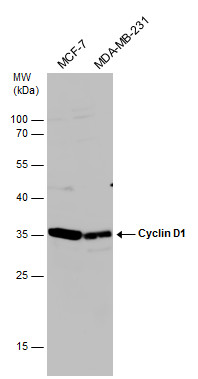
Various whole cell extracts (30 µg) were separated by 12% SDS-PAGE, and the membrane was blotted with Cyclin D1 antibody [N1C3] diluted at 1:1000. The HRP-conjugated anti-rabbit IgG antibody was used to detect the primary antibody.| Product Name | Cyclin D1 antibody |
|---|---|
| Antibody Type | Primary Antibodies |
| Product description | Rabbit Polyclonal antibody to Cyclin D1 (cyclin D1)The protein encoded by this gene belongs to the highly conserved cyclin family, whose members are characterized by a dramatic periodicity in protein abundance throughout the cell cycle. Cyclins function as regulators of CDK kinases. Different cyclins exhibit distinct expression and degradation patterns which contribute to the temporal coordination of each mitotic event. This cyclin forms a complex with and functions as a regulatory subunit of CDK4 or CDK6, whose activity is required for cell cycle G1/S transition. This protein has been shown to interact with tumor suppressor protein Rb and the expression of this gene is regulated positively by Rb. Mutations, amplification and overexpression of this gene, which alters cell cycle progression, are observed frequently in a variety of tumors and may contribute to tumorigenesis. [provided by RefSeq] |
| Immunogen | Full length human Cyclin D1 Recombinant protein. |
| Clonality | Polyclonal |
|---|---|
| Isotype | IgG |
| Host Species | Rabbit |
| Tested Applications | ICC/IFIPWB |
| *Optimal dilutions/concentrations should be determined by the researcher.
: |
|
| Species Reactivity | HumanRat |
| Predicted Reactivity | Mouse, Dog, Chicken, Rhesus Monkey, BovineMouse (93%), Dog (94%), Chicken (86%), Rhesus Monkey (99%), Bovine (93%) |
| Concentration | 1mg/ml |
| Purification | Affinity purified |
| Gene Synonyms | BCL1 antibody D11S287E antibody PRAD1 antibody U21B31 antibody CCND1 antibody B-cell CLL/lymphoma 1 antibody PRAD1 oncogene antibody G1/S-specific cyclin D1 antibody B-cell lymphoma 1 protein antibody BCL-1 oncogene antibody G1/S-specific cyclin- |
|---|---|
| Molecular Weight(MW) | 34 kDa (note) |
Application
Cyclin D1 antibody [N1C3] detects Cyclin D1 protein at cytoplasm and nucleus by immunofluorescent analysis. Sample: SK-N-SH cells were fixed in 4% paraformaldehyde at RT for 15 min. Green: Cyclin D1 protein stained by Cyclin D1 antibody [N1C3] diluted at 1:500. Red: beta Tubulin 3/ Tuj1, a cytoskeleton marker, stained by beta Tubulin 3/ Tuj1 antibody [GT11710] diluted at 1:500. Scale bar = 10 μm.
Application
Immunoprecipitation of Cyclin D1 protein from MCF-7 whole cell extracts using 5 μg of Cyclin D1 antibody [N1C3] . Western blot analysis was performed using Cyclin D1 antibody [N1C3] . EasyBlot anti-Rabbit IgG was used as a secondary reagent.
Application
Wild-type (WT) and Cyclin D1 knockout (KO) HeLa cell extracts (30 µg) were separated by 12% SDS-PAGE, and the membrane was blotted with Cyclin D1 antibody [N1C3] diluted at 1:5000. The HRP-conjugated anti-rabbit IgG antibody was used to detect the primary antibody, and the signal was developed with Trident ECL plus-Enhanced.
Application
Various whole cell extracts (30 µg) were separated by 12% SDS-PAGE, and the membrane was blotted with Cyclin D1 antibody [N1C3] diluted at 1:1000. The HRP-conjugated anti-rabbit IgG antibody was used to detect the primary antibody.| Positive Control | U87-MG , MCF-7 , MDA-MB-231 , *Huh 7 , *H184B5F5/M10 , *BT-474 , *HCC1937 , *Hs578T , *MDA-MB-231 , *MDA-MB-361 , *MDA-MB-453 , *T47D , *ZR-75-1 |
|---|---|
| Application Notes | *Optimal dilutions/concentrations should be determined by the researcher.
: |
| Form | Liquid |
|---|---|
| Storage Instructions | Keep as concentrated solution. Aliquot and store at -20ºC or below. Avoid multiple freeze-thaw cycles. |
| Storage Buffer | 1XPBS, 20% Glycerol (pH7). 0.025% ProClin 300 was added as a preservative. |


