Application
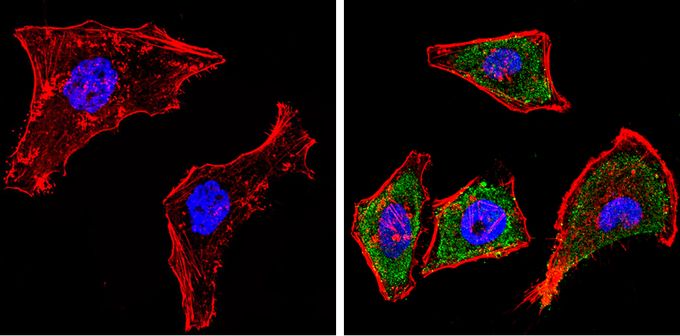
Immunofluorescent analysis of Cytochrome P450 1A1/1A2 (green) in HeLa cells. Formalin-fixed cells were permeabilized with 0.1% Triton X-100 in TBS for 5-10 minutes and blocked with 3% BSA-PBS for 30 minutes at room temperature. Cells were probed with Cytochrome P450 1A1 + 1A2 antibody [MC1] in 3% BSA-PBS at a dilution of 1:100 and incubated overnight at 4°C in a humidified chamber. Cells were washed with PBST and incubated with a proper secondary antibody. F-actin (red) was stained with a flourescent red phalloidin and nuclei (blue) were stained with Hoechst or DAPI. Images were taken at a magnification of 60x.Application
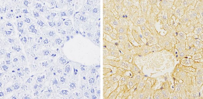
Immunohistochemistry analysis of Cytochrome P450 1A1/1A2 in paraffin-embedded rat liver tissue (right) compared with a negative control without primary antibody (left). To expose target proteins, antigen retrieval was performed using 10mM sodium citrate (pH 6.0), microwaved for 8-15 min. Following antigen retrieval, tissues were blocked in 3% H2O2-methanol for 15 min at room temperature, washed with ddH2O and PBS, and then probed with Cytochrome P450 1A1 + 1A2 antibody [MC1] diluted by 3% BSA-PBS at a dilution of 1:100 overnight at 4°C in a humidified chamber. Tissues were washed extensively in PBST and detection was performed using an HRP-conjugated secondary antibody followed by colorimetric detection using a DAB kit. Tissues were counterstained with hematoxylin and dehydrated with ethanol and xylene to prep for mounting.| Product Name | Cytochrome P450 1A1 + 1A2 antibody [MC1] |
|---|---|
| Antibody Type | Primary Antibodies |
| Product description | Mouse monoclonal antibody [MC1] to Cytochrome P450 1A1 + 1A2monooxygenase that plays a role in dioxin metabolism and detoxification which is 3-methylcholanthrene-inducible [RGD, Feb 2006] |
| Immunogen | 3-methylcholanthrene induced rat cytochrome P450 protein. |
| Clonality | Monoclonal |
|---|---|
| Isotype | IgG1 |
| Clone Number | MC1 |
| Host Species | Mouse |
| Tested Applications | ICC/IFIHC-PWB |
| IHC-P: 1:50 - 1:200 | |
| Species Reactivity | HumanMouseRatPrimate |
| Concentration | 1mg/ml |
| Gene Synonyms | cytochrome P450, family 1, subfamily a, polypeptide 1 , CP11 , Cyp45c , Cyp1a1 , P-450MC , Cypc45c , P450-C , P450DX , CYP1 , P1-450 , AHRR , AHH |
|---|---|
| Target | Cytochrome P450 1A1, 1A2 |
Application
Immunofluorescent analysis of Cytochrome P450 1A1/1A2 (green) in HeLa cells. Formalin-fixed cells were permeabilized with 0.1% Triton X-100 in TBS for 5-10 minutes and blocked with 3% BSA-PBS for 30 minutes at room temperature. Cells were probed with Cytochrome P450 1A1 + 1A2 antibody [MC1] in 3% BSA-PBS at a dilution of 1:100 and incubated overnight at 4°C in a humidified chamber. Cells were washed with PBST and incubated with a proper secondary antibody. F-actin (red) was stained with a flourescent red phalloidin and nuclei (blue) were stained with Hoechst or DAPI. Images were taken at a magnification of 60x.
Application
Immunohistochemistry analysis of Cytochrome P450 1A1/1A2 in paraffin-embedded rat liver tissue (right) compared with a negative control without primary antibody (left). To expose target proteins, antigen retrieval was performed using 10mM sodium citrate (pH 6.0), microwaved for 8-15 min. Following antigen retrieval, tissues were blocked in 3% H2O2-methanol for 15 min at room temperature, washed with ddH2O and PBS, and then probed with Cytochrome P450 1A1 + 1A2 antibody [MC1] diluted by 3% BSA-PBS at a dilution of 1:100 overnight at 4°C in a humidified chamber. Tissues were washed extensively in PBST and detection was performed using an HRP-conjugated secondary antibody followed by colorimetric detection using a DAB kit. Tissues were counterstained with hematoxylin and dehydrated with ethanol and xylene to prep for mounting.| Application Notes | IHC-P: 1:50 - 1:200 |
|---|
| Form | Liquid |
|---|---|
| Storage Instructions | -20° C, Avoid Freeze/Thaw Cycles |
| Storage Buffer | Ascites containing 0.05 % sodium azide |


