Application
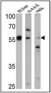
WB analysis of rat liver, H-4-II-E and HeLa lysates (25 ug) using Cytochrome P450 4A antibody at a dilution of 1:1,000.Application
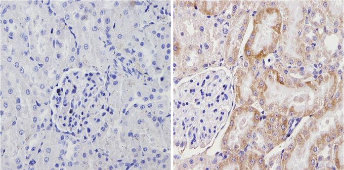
IHC-P analysis of rat kidbey tissue with (right) or without (left) Cytochrome P450 4A antibody at a dilution of 1:200. To expose target proteins, antigen retrieval method was performed using 10mM sodium citrate (pH 6.0) microwaved for 8-15 min.Application
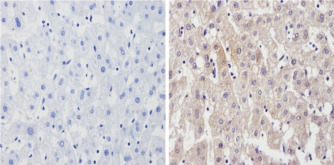
IHC-P analysis of human liver tissue with (right) or without (left) Cytochrome P450 4A antibody at a dilution of 1:200. To expose target proteins, antigen retrieval method was performed using 10mM sodium citrate (pH 6.0) microwaved for 8-15 min.Application
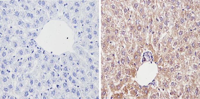
IHC-P analysis of rat liver tissue with (right) or without (left) Cytochrome P450 4A antibody at a dilution of 1:200. To expose target proteins, antigen retrieval method was performed using 10mM sodium citrate (pH 6.0) microwaved for 8-15 min.Application
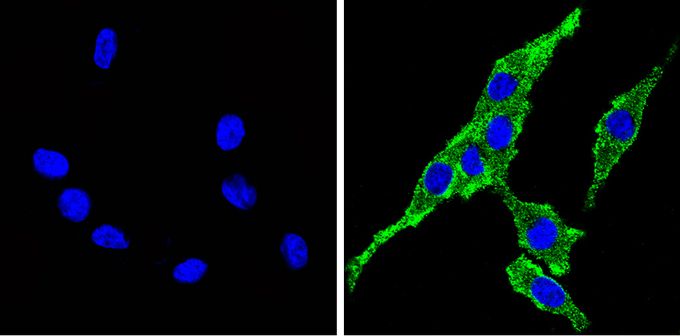
ICC/IF analysis of PC12 cells with (left) or without (right) Cytochrome P450 4A antibody at a dilution of 1:100 (green). Nuclei (blue) were stained with DAPI. Images were taken at a magnification of 60x.Application
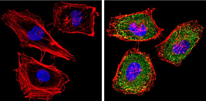
ICC/IF analysis of HeLa cells with (left) or without (right) Cytochrome P450 4A antibody at a dilution of 1:100 (green). F-actin (red) was stained with a flourescent red phalloidin and nuclei (blue) were stained with DAPI. Images were taken at a magnification of 60x.Application
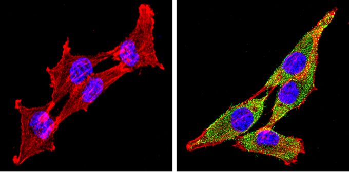
ICC/IF analysis of H-4-II-E cells with (left) or without (right) Cytochrome P450 4A antibody at a dilution of 1:100 (green). F-actin (red) was stained with a flourescent red phalloidin and nuclei (blue) were stained with DAPI. Images were taken at a magnification of 60x.| Product Name | Cytochrome P450 4A antibody |
|---|---|
| Antibody Type | Primary Antibodies |
| Product description | Rabbit Polyclonal antibody to Cytochrome P450 4AThe cytochrome P450 (P450) family of enzymes is one of three enzyme systems which metabolize the fatty acid arachadonic acid (AA) to regulators of vascular tone. P450 enzymes are monooxygenase enzymes which require several co-factors such as NADPH and P450 reductase. There are over 200 cDNA’s which encode P450 protein. Epoxygenases are those P450 proteins which metabolize AA to epoxyeicosatrienoic acid (EETs) and w-hydroxylases are those P450 proteins which produce 19- and 20-hydroxyeicosatetraenoic acids (19- and 20-HETE). 20-HETE is converted from AA by the 4A family of P450 proteins which includes at least 8 different, though closely related isoforms. 4A1, 4A2, & 4A3 have been cloned from liver, kidney and testis and have been detected in renal, hepatic & brain microvessels. |
| Immunogen | Synthetic peptide corresponding to residues D(431) P S R F A P D S P R H S H S(445) of rat Cytochrome P450 4A. |
| Clonality | Polyclonal |
|---|---|
| Host Species | Rabbit |
| Tested Applications | ICC/IFIHCIHC-FIHC-PIPWB |
| IHC-P: 1:100 - 1:500: |
|
| Species Reactivity | HumanMouseDogPigRatCatHamsterRabbit |
| Concentration | 1mg/ml |
| Purification | Affinity purified |
| Gene Synonyms | Cyp4a2 Cyp4a11 cytochrome P450 family 4 subfamily a polypeptide 2 |
|---|---|
| Target | Cytochrome P450 4A |
Application
WB analysis of rat liver, H-4-II-E and HeLa lysates (25 ug) using Cytochrome P450 4A antibody at a dilution of 1:1,000.
Application
IHC-P analysis of rat kidbey tissue with (right) or without (left) Cytochrome P450 4A antibody at a dilution of 1:200. To expose target proteins, antigen retrieval method was performed using 10mM sodium citrate (pH 6.0) microwaved for 8-15 min.
Application
IHC-P analysis of human liver tissue with (right) or without (left) Cytochrome P450 4A antibody at a dilution of 1:200. To expose target proteins, antigen retrieval method was performed using 10mM sodium citrate (pH 6.0) microwaved for 8-15 min.
Application
IHC-P analysis of rat liver tissue with (right) or without (left) Cytochrome P450 4A antibody at a dilution of 1:200. To expose target proteins, antigen retrieval method was performed using 10mM sodium citrate (pH 6.0) microwaved for 8-15 min.
Application
ICC/IF analysis of PC12 cells with (left) or without (right) Cytochrome P450 4A antibody at a dilution of 1:100 (green). Nuclei (blue) were stained with DAPI. Images were taken at a magnification of 60x.
Application
ICC/IF analysis of HeLa cells with (left) or without (right) Cytochrome P450 4A antibody at a dilution of 1:100 (green). F-actin (red) was stained with a flourescent red phalloidin and nuclei (blue) were stained with DAPI. Images were taken at a magnification of 60x.
Application
ICC/IF analysis of H-4-II-E cells with (left) or without (right) Cytochrome P450 4A antibody at a dilution of 1:100 (green). F-actin (red) was stained with a flourescent red phalloidin and nuclei (blue) were stained with DAPI. Images were taken at a magnification of 60x.| Application Notes | IHC-P: 1:100 - 1:500: |
|---|
| Form | Liquid |
|---|---|
| Storage Instructions | Keep as concentrated solution. Aliquot and store at -20ºC or below. Avoid multiple freeze-thaw cycles. |
| Storage Buffer | PBS containing 0.05% sodium azide. |


