Application
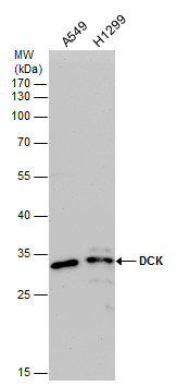
DCK antibody detects DCK protein by western blot analysis. Various whole cell extracts (30 µg) were separated by 12% SDS-PAGE, and the membrane was blotted with DCK antibody diluted at a dilution of 1:5000. The HRP-conjugated anti-rabbit IgG antibody was used to detect the primary antibody.Application
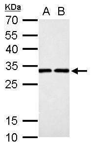
DCK antibody detects DCK protein by western blot analysis. A. 30 µg PC-12 whole cell lysate/extract B. 30 µg Rat2 whole cell lysate/extract 12% SDS-PAGE DCK antibody dilution: 1:1000 The HRP-conjugated anti-rabbit IgG antibody was used to detect the primary antibody.Application
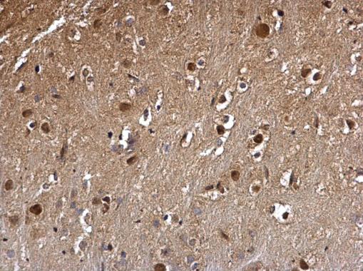
DCK antibody detects DCK protein at nucleus on mouse hind brain by immunohistochemical analysis. Sample: Paraffin-embedded mouse hind brain. DCK antibody dilution: 1:500.Application
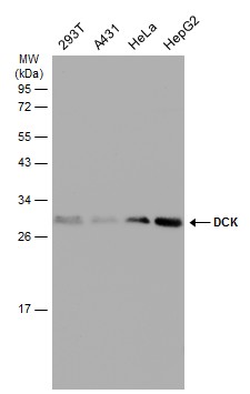
Various whole cell extracts (30 µg) were separated by 12% SDS-PAGE, and the membrane was blotted with DCK antibody diluted at 1:5000. The HRP-conjugated anti-rabbit IgG antibody was used to detect the primary antibody.Application
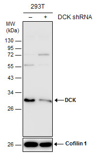
Non-transfected (–) and transfected (+) 293T whole cell extracts (30 µg) were separated by 12% SDS-PAGE, and the membrane was blotted with DCK antibody diluted at 1:10000. The HRP-conjugated anti-rabbit IgG antibody was used to detect the primary antibody.Application
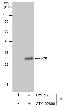
Immunoprecipitation of DCK protein from 293T whole cell extracts using 5 μg of DCK antibody . Western blot analysis was performed using DCK antibody . EasyBlot anti-Rabbit IgG was used as a secondary reagent.| Product Name | DCK antibody |
|---|---|
| Antibody Type | Primary Antibodies |
| Product description | Rabbit Polyclonal antibody to DCK (deoxycytidine kinase)Deoxycytidine kinase (DCK) is required for the phosphorylation of several deoxyribonucleosides and their nucleoside analogs. Deficiency of DCK is associated with resistance to antiviral and anticancer chemotherapeutic agents. Conversely, increased deoxycytidine kinase activity is associated with increased activation of these compounds to cytotoxic nucleoside triphosphate derivatives. DCK is clinically important because of its relationship to drug resistance and sensitivity. [provided by RefSeq] |
| Immunogen | Recombinant protein encompassing a sequence within the center region of human DCK. The exact sequence is proprietary. |
| Clonality | Polyclonal |
|---|---|
| Isotype | IgG |
| Host Species | Rabbit |
| Tested Applications | IHC-PIPWB |
| *Optimal dilutions/concentrations should be determined by the researcher.
: |
|
| Species Reactivity | HumanMouseRat |
| Predicted Reactivity | Chicken, Rhesus Monkey, BovineChicken (82%), Rhesus Monkey (96%), Bovine (95%) |
| Concentration | 1mg/ml (Please refer to the vial label for the specific concentration) |
| Purification | Affinity purified |
| Gene Synonyms | MGC117410 antibody MGC138632 antibody DCK antibody deoxycytidine kinase antibody deoxynucleoside kinase antibody |
|---|---|
| Molecular Weight(MW) | 31 kDa (note) |
| Cellular Localization | Nucleus |
Application
DCK antibody detects DCK protein by western blot analysis. Various whole cell extracts (30 µg) were separated by 12% SDS-PAGE, and the membrane was blotted with DCK antibody diluted at a dilution of 1:5000. The HRP-conjugated anti-rabbit IgG antibody was used to detect the primary antibody.
Application
DCK antibody detects DCK protein by western blot analysis. A. 30 µg PC-12 whole cell lysate/extract B. 30 µg Rat2 whole cell lysate/extract 12% SDS-PAGE DCK antibody dilution: 1:1000 The HRP-conjugated anti-rabbit IgG antibody was used to detect the primary antibody.
Application
DCK antibody detects DCK protein at nucleus on mouse hind brain by immunohistochemical analysis. Sample: Paraffin-embedded mouse hind brain. DCK antibody dilution: 1:500.
Application
Various whole cell extracts (30 µg) were separated by 12% SDS-PAGE, and the membrane was blotted with DCK antibody diluted at 1:5000. The HRP-conjugated anti-rabbit IgG antibody was used to detect the primary antibody.
Application
Non-transfected (–) and transfected (+) 293T whole cell extracts (30 µg) were separated by 12% SDS-PAGE, and the membrane was blotted with DCK antibody diluted at 1:10000. The HRP-conjugated anti-rabbit IgG antibody was used to detect the primary antibody.
Application
Immunoprecipitation of DCK protein from 293T whole cell extracts using 5 μg of DCK antibody . Western blot analysis was performed using DCK antibody . EasyBlot anti-Rabbit IgG was used as a secondary reagent.| Positive Control | 293T , A431 , HeLa , HepG2 , PC-12 , Rat2 , A549 , H1299 |
|---|---|
| Application Notes | *Optimal dilutions/concentrations should be determined by the researcher.
: |
| Form | Liquid |
|---|---|
| Storage Instructions | Keep as concentrated solution. Aliquot and store at -20ºC or below. Avoid multiple freeze-thaw cycles. |
| Storage Buffer | 1XPBS, 1% BSA, 20% Glycerol (pH7). 0.01% Thimerosal was added as a preservative. |


