Application
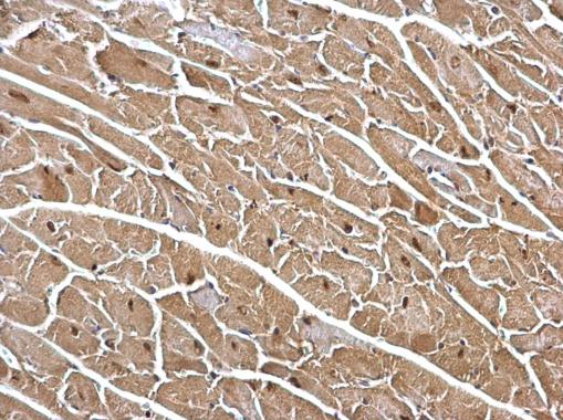
DDB1 antibody [C3], C-term detects DDB1 protein at cytosol and nucleus on mouse heart by immunohistochemical analysis. Sample: Paraffin-embedded mouse heart. DDB1 antibody [C3], C-term dilution: 1:500.Application
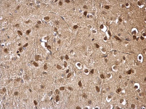
DDB1 antibody [C3], C-term detects DDB1 protein at cytosol and nucleus on mouse hind brain by immunohistochemical analysis. Sample: Paraffin-embedded mouse hind brain. DDB1 antibody [C3], C-term dilution: 1:500.Application
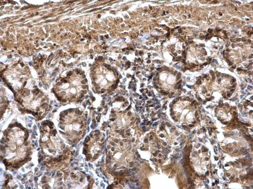
DDB1 antibody [C3], C-term detects DDB1 protein at cytosol and nucleus on mouse duodenum by immunohistochemical analysis. Sample: Paraffin-embedded mouse duodenum. DDB1 antibody [C3], C-term dilution: 1:500.Application
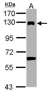
Sample (30 µg of whole cell lysate) A: PC-12 7.5% SDS PAGE diluted at 1:1000 The HRP-conjugated anti-rabbit IgG antibody was used to detect the primary antibody.Application
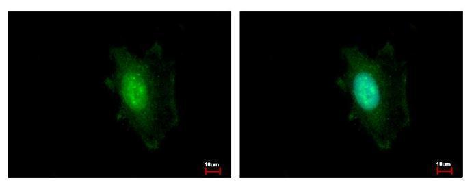
DDB1 antibody [C3], C-term detects DDB1 protein at cytoplasm and nucleus by immunofluorescent analysis. Sample: HeLa cells were fixed in ice-cold MeOH for 5 min. Green: DDB1 protein stained by DDB1 antibody [C3], C-term diluted at 1:500. Blue: Hoechst 33343 staining.Application
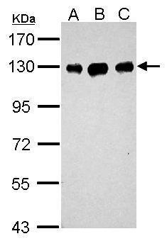
Sample (30 µg of whole cell lysate) A: Neuro2A B: GL261 C: C8D30 7.5% SDS PAGE diluted at 1:1000 The HRP-conjugated anti-rabbit IgG antibody was used to detect the primary antibody.Application
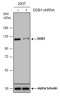
Non-transfected (–) and transfected (+) 293T whole cell extracts (30 µg) were separated by 7.5% SDS-PAGE, and the membrane was blotted with DDB1 antibody [C3], C-term diluted at 1:10000. The HRP-conjugated anti-rabbit IgG antibody was used to detect the primary antibody.Application
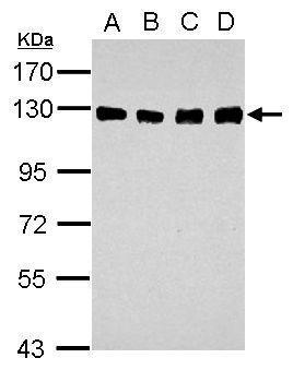
Sample (30 µg of whole cell lysate) A: A549 B: H1299 C: HCT116 D: MCF-7 7.5% SDS PAGE diluted at 1:1000 The HRP-conjugated anti-rabbit IgG antibody was used to detect the primary antibody.Application
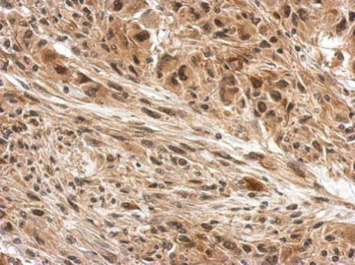
Immunohistochemical analysis of paraffin-embedded U373 xenograft, using DDB1 antibody at 1:500 dilution.| Product Name | DDB1 antibody , C-term |
|---|---|
| Antibody Type | Primary Antibodies |
| Product description | Rabbit Polyclonal antibody to DDB1 (damage-specific DNA binding protein 1, 127kDa)This gene encodes the large subunit of DNA damage-binding protein which is a heterodimer composed of a large and a small subunit. This protein functions in nucleotide-excision repair. Its defective activity causes the repair defect in the patients with xeroderma pigmentosum complementation group E (XPE). However, it remains for mutation analysis to demonstrate whether the defect in XPE patients is in this gene or the gene encoding the small subunit. In addition, Best vitelliform mascular dystrophy is mapped to the same region as this gene on 11q, but no sequence alternations of this gene are demonstrated in Best disease patients. [provided by RefSeq] |
| Immunogen | Carrier-protein conjugated synthetic peptide encompassing a sequence within the C-terminus region of human DDB1. The exact sequence is proprietary. |
| Clonality | Polyclonal |
|---|---|
| Isotype | IgG |
| Host Species | Rabbit |
| Tested Applications | ICC/IFIHC-PWB |
| *Optimal dilutions/concentrations should be determined by the researcher.
: |
|
| Species Reactivity | HumanMouseRat |
| Predicted Reactivity | Xenopus laevis, BovineXenopus laevis (100%), Bovine (100%) |
| Concentration | 1mg/ml (Please refer to the vial label for the specific concentration) |
| Purification | Affinity purified |
| Gene Synonyms | DDBA antibody UV-DDB1 antibody XAP1 antibody XPCE antibody XPE antibody XPE-BF antibody DDB1 antibody DNA damage-binding protein a antibody XPE-binding factor antibody UV-DDB 1 antibody xeroderma pigmentosum group E-complementing protein antibody |
|---|---|
| Molecular Weight(MW) | 127 kDa (note) |
| Cellular Localization | Cytoplasm , Nucleus |
Application
DDB1 antibody [C3], C-term detects DDB1 protein at cytosol and nucleus on mouse heart by immunohistochemical analysis. Sample: Paraffin-embedded mouse heart. DDB1 antibody [C3], C-term dilution: 1:500.
Application
DDB1 antibody [C3], C-term detects DDB1 protein at cytosol and nucleus on mouse hind brain by immunohistochemical analysis. Sample: Paraffin-embedded mouse hind brain. DDB1 antibody [C3], C-term dilution: 1:500.
Application
DDB1 antibody [C3], C-term detects DDB1 protein at cytosol and nucleus on mouse duodenum by immunohistochemical analysis. Sample: Paraffin-embedded mouse duodenum. DDB1 antibody [C3], C-term dilution: 1:500.
Application
Sample (30 µg of whole cell lysate) A: PC-12 7.5% SDS PAGE diluted at 1:1000 The HRP-conjugated anti-rabbit IgG antibody was used to detect the primary antibody.
Application
DDB1 antibody [C3], C-term detects DDB1 protein at cytoplasm and nucleus by immunofluorescent analysis. Sample: HeLa cells were fixed in ice-cold MeOH for 5 min. Green: DDB1 protein stained by DDB1 antibody [C3], C-term diluted at 1:500. Blue: Hoechst 33343 staining.
Application
Sample (30 µg of whole cell lysate) A: Neuro2A B: GL261 C: C8D30 7.5% SDS PAGE diluted at 1:1000 The HRP-conjugated anti-rabbit IgG antibody was used to detect the primary antibody.
Application
Non-transfected (–) and transfected (+) 293T whole cell extracts (30 µg) were separated by 7.5% SDS-PAGE, and the membrane was blotted with DDB1 antibody [C3], C-term diluted at 1:10000. The HRP-conjugated anti-rabbit IgG antibody was used to detect the primary antibody.
Application
Sample (30 µg of whole cell lysate) A: A549 B: H1299 C: HCT116 D: MCF-7 7.5% SDS PAGE diluted at 1:1000 The HRP-conjugated anti-rabbit IgG antibody was used to detect the primary antibody.
Application
Immunohistochemical analysis of paraffin-embedded U373 xenograft, using DDB1 antibody at 1:500 dilution.| Positive Control | 293T , A549 , H1299 , HCT116 , MCF-7 , Neuro2A , GL261 , C8D30 , PC-12 |
|---|---|
| Application Notes | *Optimal dilutions/concentrations should be determined by the researcher.
: |
| Form | Liquid |
|---|---|
| Storage Instructions | Keep as concentrated solution. Aliquot and store at -20ºC or below. Avoid multiple freeze-thaw cycles. |
| Storage Buffer | 1XPBS, 20% Glycerol (pH7). 0.01% Thimerosal was added as a preservative. |


