Application
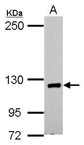
Sample (30 µg of whole cell lysate) A: 293T 5% SDS PAGE diluted at 1:1000 The HRP-conjugated anti-rabbit IgG antibody was used to detect the primary antibody.Application
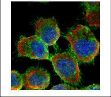
Confocal immunofluorescence analysis (Olympus FV10i) of methanol-fixed A431, using E-cadherin antibody (Green) at 1:500 dilution. Alpha-tubulin filaments were labeled with (Red) at 1:2000.Application
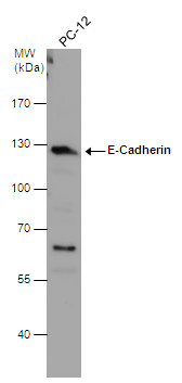
E-Cadherin antibody [N3C2], Internal detects E-Cadherin protein by western blot analysis. Whole cell extracts (30 µg) was separated by 7.5% SDS-PAGE, and the membrane was blotted with E-Cadherin antibody [N3C2], Internal diluted at 1:3000. The HRP-conjugated anti-rabbit IgG antibody was used to detect the primary antibody.Application
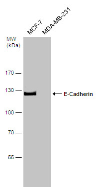
Various whole cell extracts (30 µg) were separated by 7.5% SDS-PAGE, and the membrane was blotted with E-Cadherin antibody [N3C2], Internal diluted at 1:2000. The HRP-conjugated anti-rabbit IgG antibody was used to detect the primary antibody.Application
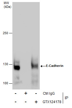
Immunoprecipitation of E-Cadherin protein from MCF-7 whole cell extracts using 5 μg of E-Cadherin antibody [N3C2], Internal . Western blot analysis was performed using E-Cadherin antibody [N3C2], Internal . EasyBlot anti-Rabbit IgG was used as a secondary reagent.Application
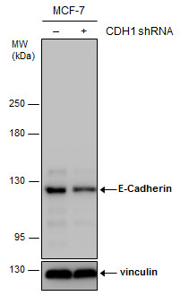
Non-transfected (–) and transfected (+) MCF-7 whole cell extracts (30 µg) were separated by 5% SDS-PAGE, and the membrane was blotted with E-Cadherin antibody [N3C2], Internal diluted at 1:1000. The HRP-conjugated anti-rabbit IgG antibody was used to detect the primary antibody.| Product Name | E-Cadherin antibody , Internal |
|---|---|
| Antibody Type | Primary Antibodies |
| Product description | Rabbit Polyclonal antibody to E-Cadherin (cadherin 1, type 1, E-cadherin (epithelial))This gene is a classical cadherin from the cadherin superfamily. The encoded protein is a calcium dependent cell-cell adhesion glycoprotein comprised of five extracellular cadherin repeats, a transmembrane region and a highly conserved cytoplasmic tail. Mutations in this gene are correlated with gastric, breast, colorectal, thyroid and ovarian cancer. Loss of function is thought to contribute to progression in cancer by increasing proliferation, invasion, and/or metastasis. The ectodomain of this protein mediates bacterial adhesion to mammalian cells and the cytoplasmic domain is required for internalization. Identified transcript variants arise from mutation at consensus splice sites. [provided by RefSeq] |
| Immunogen | Recombinant protein encompassing a sequence within the center region of human E-Cadherin. The exact sequence is proprietary. |
| Modification | Acetylated |
| Clonality | Polyclonal |
|---|---|
| Isotype | IgG |
| Host Species | Rabbit |
| Tested Applications | ICC/IFIPWB |
| WB:1:500~1:2000 IP:1:100~1:500 ICC/IF:1:100~1:500 *Optimal dilutions/concentrations should be determined by the researcher.: |
|
| Species Reactivity | HumanRat |
| Predicted Reactivity | Pig, BovinePig (82%), Bovine (83%) |
| Concentration | 1mg/ml (Please refer to the vial label for the specific concentration) |
| Purification | Affinity purified |
| Gene Synonyms | Arc-1 antibody CD324 antibody CDHE antibody ECAD antibody LCAM antibody UVO antibody CDH1 antibody epithelial cadherin antibody |
|---|---|
| Molecular Weight(MW) | 97 kDa (note) (2) |
| Cellular Localization | Cell junction , Cell membrane |
Application
Sample (30 µg of whole cell lysate) A: 293T 5% SDS PAGE diluted at 1:1000 The HRP-conjugated anti-rabbit IgG antibody was used to detect the primary antibody.
Application
Confocal immunofluorescence analysis (Olympus FV10i) of methanol-fixed A431, using E-cadherin antibody (Green) at 1:500 dilution. Alpha-tubulin filaments were labeled with (Red) at 1:2000.
Application
E-Cadherin antibody [N3C2], Internal detects E-Cadherin protein by western blot analysis. Whole cell extracts (30 µg) was separated by 7.5% SDS-PAGE, and the membrane was blotted with E-Cadherin antibody [N3C2], Internal diluted at 1:3000. The HRP-conjugated anti-rabbit IgG antibody was used to detect the primary antibody.
Application
Various whole cell extracts (30 µg) were separated by 7.5% SDS-PAGE, and the membrane was blotted with E-Cadherin antibody [N3C2], Internal diluted at 1:2000. The HRP-conjugated anti-rabbit IgG antibody was used to detect the primary antibody.
Application
Immunoprecipitation of E-Cadherin protein from MCF-7 whole cell extracts using 5 μg of E-Cadherin antibody [N3C2], Internal . Western blot analysis was performed using E-Cadherin antibody [N3C2], Internal . EasyBlot anti-Rabbit IgG was used as a secondary reagent.
Application
Non-transfected (–) and transfected (+) MCF-7 whole cell extracts (30 µg) were separated by 5% SDS-PAGE, and the membrane was blotted with E-Cadherin antibody [N3C2], Internal diluted at 1:1000. The HRP-conjugated anti-rabbit IgG antibody was used to detect the primary antibody.| Positive Control | 293T , PC-12 , MCF-7 |
|---|---|
| Application Notes | WB:1:500~1:2000 IP:1:100~1:500 ICC/IF:1:100~1:500 *Optimal dilutions/concentrations should be determined by the researcher.: |
| Form | Liquid |
|---|---|
| Storage Instructions | Keep as concentrated solution. Aliquot and store at -20ºC or below. Avoid multiple freeze-thaw cycles. |
| Storage Buffer | 1XPBS, 20% Glycerol (pH7). 0.01% Thimerosal was added as a preservative. |


