Application
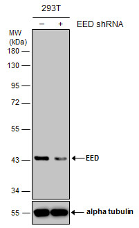
WB ImageNon-transfected (–) and transfected (+) 293T whole cell extracts (30 µg) were separated by 10% SDS-PAGE, and the membrane was blotted with EED antibody diluted at 1:500.Application
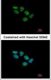
ICC/IF ImageImmunofluorescence analysis of paraformaldehyde-fixed A431, using EED antibody at 1:500 dilution.Application
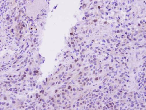
IHC-P ImageImmunohistochemical analysis of paraffin-embedded SAS Xenograft, using EED antibody at 1:100 dilution.Application
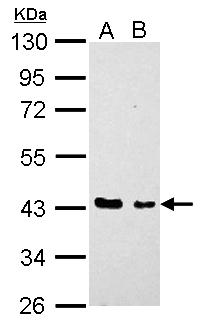
WB ImageEED antibody detects EED protein by Western blot analysis. A. 30 µg H1299 whole cell lysate/extract B. 30 µg HCT116 whole cell lysate/extract 10 % SDS-PAGE EED antibody dilution: 1:1000| Product Name | EED antibody |
|---|---|
| Antibody Type | Primary Antibodies |
| Product description | Rabbit Polyclonal antibody to EED (embryonic ectoderm development)This gene encodes a member of the Polycomb-group (PcG) family. PcG family members form multimeric protein complexes, which are involved in maintaining the transcriptional repressive state of genes over successive cell generations. This protein interacts with enhancer of zeste 2, the cytoplasmic tail of integrin beta7, immunodeficiency virus type 1 (HIV-1) MA protein, and histone deacetylase proteins. This protein mediates repression of gene activity through histone deacetylation, and may act as a specific regulator of integrin function. Two transcript variants encoding distinct isoforms have been identified for this gene. [provided by RefSeq] |
| Immunogen | Recombinant protein encompassing a sequence within the center region of human EED. The exact sequence is proprietary. |
| Clonality | Polyclonal |
|---|---|
| Isotype | IgG |
| Host Species | Rabbit |
| Tested Applications | ICC/IFIHC-PWB |
| *Optimal dilutions/concentrations should be determined by the researcher.
: |
|
| Species Reactivity | Human |
| Concentration | 1mg/ml (Please refer to the vial label for the specific concentration) |
| Purification | Affinity purified |
| Gene Synonyms | HEED antibody WAIT1 antibody EED antibody WD protein associating with integrin cytoplasmic tails 1 antibody polycomb protein EED antibody embryonic ectoderm development antibody |
|---|---|
| Molecular Weight(MW) | 50 kDa (note) (4) |
Application
WB ImageNon-transfected (–) and transfected (+) 293T whole cell extracts (30 µg) were separated by 10% SDS-PAGE, and the membrane was blotted with EED antibody diluted at 1:500.
Application
ICC/IF ImageImmunofluorescence analysis of paraformaldehyde-fixed A431, using EED antibody at 1:500 dilution.
Application
IHC-P ImageImmunohistochemical analysis of paraffin-embedded SAS Xenograft, using EED antibody at 1:100 dilution.
Application
WB ImageEED antibody detects EED protein by Western blot analysis. A. 30 µg H1299 whole cell lysate/extract B. 30 µg HCT116 whole cell lysate/extract 10 % SDS-PAGE EED antibody dilution: 1:1000| Application Notes | *Optimal dilutions/concentrations should be determined by the researcher.
: |
|---|
| Form | Liquid |
|---|---|
| Storage Instructions | Keep as concentrated solution. Aliquot and store at -20ºC or below. Avoid multiple freeze-thaw cycles. |
| Storage Buffer | 1XPBS, 20% Glycerol (pH7). 0.01% Thimerosal was added as a preservative. |


