Application
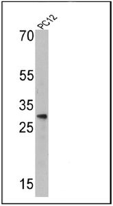
Western blot analysis of Endothelin 1 in 25 ug of PC12 cell lysates. Proteins were transferred to a PVDF membrane and blocked at 4°C overnight. The membrane was probed with Endothelin 1 antibody [TR.ET.48.5] at a dilution of 1:500 overnight at 4°C, washed in TBST, and probed with an HRP-conjugated secondary antibody for 1 hr at room temperature in the dark. Chemiluminescent detection was performed.Application
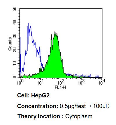
Flow cytometry analysis of Endothelin 1 in HepG2 cells compared to an isotype control (blue). Cells were harvested, adjusted to a concentration of 1-5x10^6 cells/ml, fixed with 2% paraformaldehyde, washed with PBS, and incubated with Endothelin 1 antibody [TR.ET.48.5] at a dilution of 0.5 ug/test for 60 min at room temperature. Cells were then blocked in a solution of 2% BSA-PBS for 30 min at room temperature, incubated for 40 min at room temperature in the dark using a proper secondary antibody, and re-suspended in PBS for FACS analysis.Application
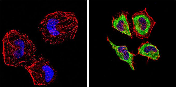
Immunofluorescent analysis of Endothelin 1 (green) in HeLa cells compared with a negative control in the absence of primary antibody (left). Formalin-fixed cells were permeabilized with 0.1% Triton X-100 in TBS for 5-10 minutes, blocked with 3% BSA-PBS for 30 minutes at room temperature and probed with Endothelin 1 antibody [TR.ET.48.5] in 3% BSA-PBS at a dilution of 1:200 and incubated overnight at 4°C in a humidified chamber. Cells were washed with PBST and incubated with a proper secondary antibody in PBS at room temperature in the dark. F-actin (red) was stained with a flourescent red phalloidin and nuclei (blue) were stained with DAPI for 5-10 minutes in the dark. Images were taken at a magnification of 60x.| Product Name | Endothelin 1 antibody [TR.ET.48.5] |
|---|---|
| Antibody Type | Primary Antibodies |
| Product description | Mouse monoclonal antibody [TR.ET.48.5] to Endothelin 1The protein encoded by this gene is proteolytically processed to release a secreted peptide termed endothelin 1. This peptide is a potent vasoconstrictor and is produced by vascular endothelial cells. Endothelin 1 also can affect the central nervous system. Two transcript variants encoding different isoforms have been found for this gene. [provided by RefSeq, Nov 2009] |
| Immunogen | The immunogen corresponding to a region within amino acids 60 and 68 of human ET-1 (Uniprot: P05305). |
| Clonality | Monoclonal |
|---|---|
| Isotype | IgG1 |
| Clone Number | TR.ET.48.5 |
| Host Species | Mouse |
| Tested Applications | BlockingELISAFACSICC/IFIHCIHC-FIHC-PIPRIAWB |
| ICC/IF: 1:200 - 1:1000 | |
| Species Reactivity | HumanMouseDogPigRatRabbitSheep |
| Concentration | 1mg/ml |
| Purification | Affinity purified |
| Gene Synonyms | ET1 , EDN1 , endothelin 1 , HDLCQ7 , PPET1 |
|---|---|
| Target | Endothelin 1 |
| Molecular Weight(MW) | 24 kDa (note) |
| Tissue Specificity | This antibody shows little cross-reactivity to related peptides and no known cross-reactivity to non-related peptides. This antibody is not recommended for human placenta tissue or HepG2 cells in Western blot applications. |
Application
Western blot analysis of Endothelin 1 in 25 ug of PC12 cell lysates. Proteins were transferred to a PVDF membrane and blocked at 4°C overnight. The membrane was probed with Endothelin 1 antibody [TR.ET.48.5] at a dilution of 1:500 overnight at 4°C, washed in TBST, and probed with an HRP-conjugated secondary antibody for 1 hr at room temperature in the dark. Chemiluminescent detection was performed.
Application
Flow cytometry analysis of Endothelin 1 in HepG2 cells compared to an isotype control (blue). Cells were harvested, adjusted to a concentration of 1-5x10^6 cells/ml, fixed with 2% paraformaldehyde, washed with PBS, and incubated with Endothelin 1 antibody [TR.ET.48.5] at a dilution of 0.5 ug/test for 60 min at room temperature. Cells were then blocked in a solution of 2% BSA-PBS for 30 min at room temperature, incubated for 40 min at room temperature in the dark using a proper secondary antibody, and re-suspended in PBS for FACS analysis.
Application
Immunofluorescent analysis of Endothelin 1 (green) in HeLa cells compared with a negative control in the absence of primary antibody (left). Formalin-fixed cells were permeabilized with 0.1% Triton X-100 in TBS for 5-10 minutes, blocked with 3% BSA-PBS for 30 minutes at room temperature and probed with Endothelin 1 antibody [TR.ET.48.5] in 3% BSA-PBS at a dilution of 1:200 and incubated overnight at 4°C in a humidified chamber. Cells were washed with PBST and incubated with a proper secondary antibody in PBS at room temperature in the dark. F-actin (red) was stained with a flourescent red phalloidin and nuclei (blue) were stained with DAPI for 5-10 minutes in the dark. Images were taken at a magnification of 60x.| Application Notes | ICC/IF: 1:200 - 1:1000 |
|---|
| Form | Liquid |
|---|---|
| Storage Instructions | -20° C, Avoid Freeze/Thaw Cycles |
| Storage Buffer | PBS containing 0.05 % sodium azide |


