Application
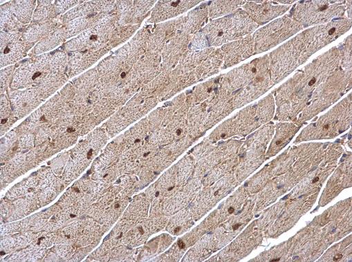
ERR alpha antibody [N1], N-term detects ERR alpha protein at nucleus on mouse heart by immunohistochemical analysis. Sample: Paraffin-embedded mouse heart. ERR alpha antibody [N1], N-term diluted at 1:500.Application
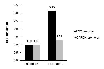
Cross-linked ChIP was performed with MCF-7 chromatin extract and 5 μg of either control rabbit IgG or anti-ERR alpha antibody. The precipitated DNA was detected by PCR with primer set targeting to PS2 promoter or GAPDH promoter.Application
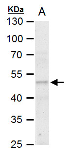
ERR alpha antibody [N1], N-term detects ERR alpha protein by western blot analysis. A. 50 µg rat kideny extract 10% SDS-PAGE ERR alpha antibody [N1], N-term dilution: 1:500 The HRP-conjugated anti-rabbit IgG antibody was used to detect the primary antibody.Application
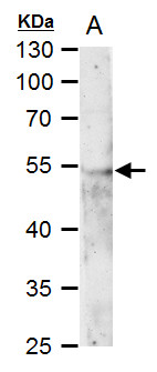
ERR alpha antibody [N1], N-term detects ERR alpha protein by western blot analysis. A. 50 µg mouse kideny extract 10% SDS-PAGE ERR alpha antibody [N1], N-term dilution: 1:500 The HRP-conjugated anti-rabbit IgG antibody was used to detect the primary antibody.Application
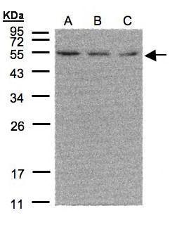
Sample(30 µg of whole cell lysate) A:293T B:A431 C:H1299 12% SDS PAGE diluted at 1:500 The HRP-conjugated anti-rabbit IgG antibody was used to detect the primary antibody.Application
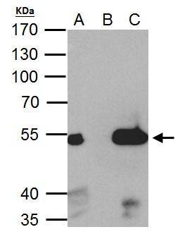
ERR alpha antibody immunoprecipitates ERR alpha protein in IP experiments. IP Sample: 293T whole cell lysate/extract A. 40 μg 293T whole cell lysate/extract B. Control with 2 μg of preimmune rabbit IgG C. Immunoprecipitation of ERR alpha protein by 2 μg of ERR alpha antibody 7.5% SDS-PAGE The immunoprecipitated ERR alpha protein was detected by ERR alpha antibody diluted at 1:1000. EasyBlot anti-rabbit IgG was used as a secondary reagent.| Product Name | ERR alpha antibody , N-term |
|---|---|
| Antibody Type | Primary Antibodies |
| Product description | Rabbit Polyclonal antibody to ERR alpha (estrogen-related receptor alpha)The protein encoded by this gene is a nuclear receptor that is closely related to the estrogen receptor. This protein acts as a site-specific transcription regulator and has been also shown to interact with estrogen and the transcripton factor TFIIB by direct protein-protein contact. The binding and regulatory activities of this protein have been demonstrated in the regulation of a variety of genes including lactoferrin, osteopontin, medium-chain acyl coenzyme A dehydrogenase (MCAD) and thyroid hormone receptor genes. A processed pseudogene of ESRRA is located on chromosome 13q12.1. [provided by RefSeq] |
| Immunogen | Carrier-protein conjugated synthetic peptide encompassing a sequence within the N-terminus region of human ERR alpha. The exact sequence is proprietary. |
| Clonality | Polyclonal |
|---|---|
| Isotype | IgG |
| Host Species | Rabbit |
| Tested Applications | IHC-PIPWBChIP |
| *Optimal dilutions/concentrations should be determined by the researcher.
: |
|
| Species Reactivity | HumanMouseRat |
| Predicted Reactivity | Dog, Pig, BovineDog (100%), Pig (100%), Bovine (100%) |
| Concentration | 1mg/ml |
| Purification | Affinity purified |
| Gene Synonyms | ERR1 antibody ERRa antibody ERRalpha antibody ESRL1 antibody NR3B1 antibody ESRRA antibody estrogen-related nuclear receptor alpha antibody estrogen receptor-like 1 antibody steroid hormone receptor ERR1 antibody nuclear receptor subfamily 3 group |
|---|---|
| Molecular Weight(MW) | 46 kDa (note) (3) |
| Cellular Localization | Nucleus |
Application
ERR alpha antibody [N1], N-term detects ERR alpha protein at nucleus on mouse heart by immunohistochemical analysis. Sample: Paraffin-embedded mouse heart. ERR alpha antibody [N1], N-term diluted at 1:500.
Application
Cross-linked ChIP was performed with MCF-7 chromatin extract and 5 μg of either control rabbit IgG or anti-ERR alpha antibody. The precipitated DNA was detected by PCR with primer set targeting to PS2 promoter or GAPDH promoter.
Application
ERR alpha antibody [N1], N-term detects ERR alpha protein by western blot analysis. A. 50 µg rat kideny extract 10% SDS-PAGE ERR alpha antibody [N1], N-term dilution: 1:500 The HRP-conjugated anti-rabbit IgG antibody was used to detect the primary antibody.
Application
ERR alpha antibody [N1], N-term detects ERR alpha protein by western blot analysis. A. 50 µg mouse kideny extract 10% SDS-PAGE ERR alpha antibody [N1], N-term dilution: 1:500 The HRP-conjugated anti-rabbit IgG antibody was used to detect the primary antibody.
Application
Sample(30 µg of whole cell lysate) A:293T B:A431 C:H1299 12% SDS PAGE diluted at 1:500 The HRP-conjugated anti-rabbit IgG antibody was used to detect the primary antibody.
Application
ERR alpha antibody immunoprecipitates ERR alpha protein in IP experiments. IP Sample: 293T whole cell lysate/extract A. 40 μg 293T whole cell lysate/extract B. Control with 2 μg of preimmune rabbit IgG C. Immunoprecipitation of ERR alpha protein by 2 μg of ERR alpha antibody 7.5% SDS-PAGE The immunoprecipitated ERR alpha protein was detected by ERR alpha antibody diluted at 1:1000. EasyBlot anti-rabbit IgG was used as a secondary reagent.| Positive Control | 293T , A431 , H1299 , Mouse kidney , Rat kidney , *A549 |
|---|---|
| Application Notes | *Optimal dilutions/concentrations should be determined by the researcher.
: |
| Form | Liquid |
|---|---|
| Storage Instructions | Keep as concentrated solution. Aliquot and store at -20ºC or below. Avoid multiple freeze-thaw cycles. |
| Storage Buffer | 0.1M Tris, 0.1M Glycine, 10% Glycerol (pH7). 0.01% Thimerosal was added as a preservative. |


