
Application
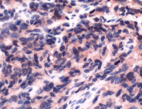
Human Ewing Sarcoma stained with anti-FLI-1 antibodyApplication
at 1/50 dilution, staining FLI1 in angiosarcoma by Immunohistochemistry (FFPE-sections)| Product Name | FLI1 antibody |
|---|---|
| Antibody Type | Primary Antibodies |
| Product description | Rabbit polyclonal to FLI1FLI1 is a member of ETS family of transcription factors and is a proto-oncogene involved in erythroleukemia and Ewings sarcoma. FLI1 was originally identified as a common proviral insertion (Friend leukemia insertion site 1) and found in 75% of erythroleukemia cell clones induced by the Friend mouse leukemia virus. A chromosomal translocation between the FLI1 gene and EWS gene is found in most Ewing sarcomas. FLI1 has anti-apoptotic acivities and also interferes with nuclear hormone receptors. |
| Immunogen | Synthetic peptide – Immunogen sequence information is commercially sensitive. The only information available is the it belongs to the C-terminus. |
| Clonality | Polyclonal |
|---|---|
| Isotype | IgG |
| Host Species | Rabbit |
| Tested Applications | IHC-P |
| For IHC-P: Use at a dilution of 1:50. Perform enzymatic antigen retrieval before commencing with IHC staining protocol, by boiling tissue sections in 10mM citrate buffer, pH 6.0 for 10 min followed by cooling at RT for 20 min. For IP: Use at a concentration of 2 μg/mg protein lysate. For WB: Use at a concentration of 2 μg/ml for 2hrs. Predicted molecular weight: 54 kDa. Optimal dilutions/concentrations should be determined by the end user.: |
|
| Species Reactivity | Human |
| Concentration | 1mg/ml |
| Purification | Affinity purified |
| Gene Synonyms | Q01543 SIC1 SIC-1 FLI1 EWSR2 2313 193067 SIC 1 |
|---|---|
| Target | FLI1 |
| Cellular Localization | Nuclear |
Application
Human Ewing Sarcoma stained with anti-FLI-1 antibody
Application
at 1/50 dilution, staining FLI1 in angiosarcoma by Immunohistochemistry (FFPE-sections)| Positive Control | Angiosarcoma |
|---|---|
| Application Notes | For IHC-P: Use at a dilution of 1:50. Perform enzymatic antigen retrieval before commencing with IHC staining protocol, by boiling tissue sections in 10mM citrate buffer, pH 6.0 for 10 min followed by cooling at RT for 20 min. For IP: Use at a concentration of 2 μg/mg protein lysate. For WB: Use at a concentration of 2 μg/ml for 2hrs. Predicted molecular weight: 54 kDa. Optimal dilutions/concentrations should be determined by the end user.: |
| Form | Liquid |
|---|---|
| Storage Instructions | Keep as concentrated solution, aliquot and store at 4°C. Do not freeze. |
| Storage Buffer | PBS/1% BSA buffer pH 7.6 with less than 0.1% sodium azide |


