Application
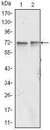
Western blot analysis using FMR1 mAb against Jurkat (1) and K562 (2) cell lysate.Application
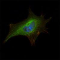
Immunofluorescence analysis of NIH-3T3 cells using FMR1 mAb (green). Blue: DRAQ5 fluorescent DNA dye. Red: Actin filaments have been labeled with Alexa Fluor-555 phalloidin.Application
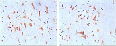
Immunohistochemical analysis of paraffin-embedded human brain tissues, showing cytoplasmic localization with DAB staining using FMR1 mAb.| Product Name | FMRP antibody |
|---|---|
| Antibody Type | Primary Antibodies |
| Product description | Mouse monoclonal antibody [4G9] to FMRPThe protein encoded by this gene binds RNA and is associated with polysomes. The encoded protein may be involved in mRNA trafficking from the nucleus to the cytoplasm. A trinucleotide repeat (CGG) in the 5' UTR is normally found at 6-53 copies, but an expansion to 55-230 repeats is the cause of fragile X syndrome. Expansion of the trinucleotide repeat may also cause one form of premature ovarian failure (POF1). Multiple alternatively spliced transcript variants that encode different protein isoforms and which are located in different cellular locations have been described for this gene. [provided by RefSeq, May 2010] |
| Immunogen | The immunogen corresponding to human FMRP expressed in E. Coli. |
| Clonality | Monoclonal |
|---|---|
| Isotype | IgG1 |
| Clone Number | 4G9 |
| Host Species | Mouse |
| Tested Applications | ELISAICC/IFIHCIHC-PWB |
| ELISA: 1/10000.: |
|
| Species Reactivity | Human |
| Concentration | 1mg/ml |
| Gene Synonyms | POF1 POF FMRP FRAXA |
|---|---|
| Target | FMRP |
| Molecular Weight(MW) | 71 kDa (note) |
Application
Western blot analysis using FMR1 mAb against Jurkat (1) and K562 (2) cell lysate.
Application
Immunofluorescence analysis of NIH-3T3 cells using FMR1 mAb (green). Blue: DRAQ5 fluorescent DNA dye. Red: Actin filaments have been labeled with Alexa Fluor-555 phalloidin.
Application
Immunohistochemical analysis of paraffin-embedded human brain tissues, showing cytoplasmic localization with DAB staining using FMR1 mAb.| Application Notes | ELISA: 1/10000.: |
|---|
| Form | Liquid |
|---|---|
| Storage Instructions | Store at 4℃, for long term storage, store at -20℃. |
| Storage Buffer | Ascitic fluid containing 0.03% sodium azide. |


