Application
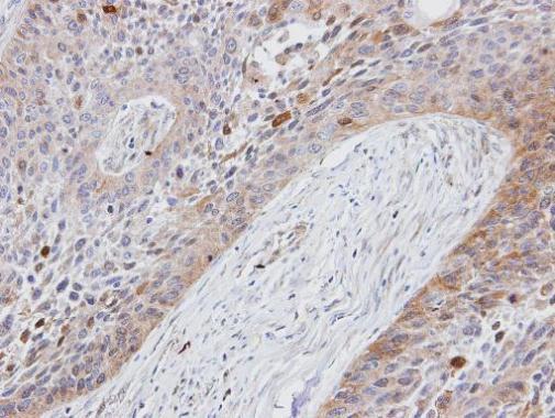
IHC-P ImageImmunohistochemical analysis of paraffin-embedded human lung cancer, using Folate Receptor alpha antibody at 1:100 dilution.Application
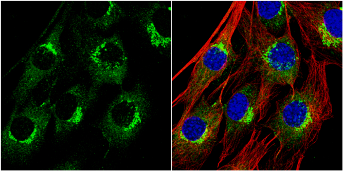
ICC/IF ImageFolate Receptor alpha antibody [N1C3] detects Folate Receptor alpha protein at cytoplasm by immunofluorescent analysis. Sample: C2C12 cells were fixed in 4% paraformaldehyde at RT for 15 min. Green: Folate Receptor alpha protein stained by Folate Receptor alpha antibody [N1C3] diluted at 1:200. Red: alpha Tubulin, a cytoskeleton marker, stained by alpha Tubulin antibody [GT114] diluted at 1:1000. Blue: Hoechst 33342 staining.Application
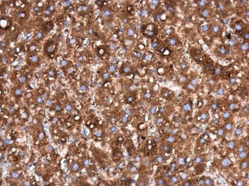
IHC-P ImageFolate Receptor alpha antibody [N1C3] detects Folate Receptor alpha protein at cytoplasm in rat liver by immunohistochemical analysis. Sample: Paraffin-embedded rat liver. Folate Receptor alpha antibody [N1C3] diluted at 1:200.| Product Name | Folate Receptor alpha antibody |
|---|---|
| Antibody Type | Primary Antibodies |
| Product description | Rabbit Polyclonal antibody to Folate Receptor alpha (folate receptor 1 (adult))The protein encoded by this gene is a member of the folate receptor (FOLR) family. Members of this gene family have a high affinity for folic acid and for several reduced folic acid derivatives, and mediate delivery of 5-methyltetrahydrofolate to the interior of cells. This gene is composed of 7 exons; exons 1 through 4 encode the 5' UTR and exons 4 through 7 encode the open reading frame. Due to the presence of 2 promoters, multiple transcription start sites, and alternative splicing of exons, several transcript variants are derived from this gene. These variants differ in the lengths of 5' and 3' UTR, but they encode an identical amino acid sequence. [provided by RefSeq] |
| Immunogen | Recombinant protein encompassing a sequence within the center region of human Folate Receptor alpha. The exact sequence is proprietary. |
| Modification | Acetylated |
| Clonality | Polyclonal |
|---|---|
| Isotype | IgG |
| Host Species | Rabbit |
| Tested Applications | ICC/IFIHC-PWB |
| WB:1:500~1:2000 ICC/IF:1:50~1:100 IHC-p:1:50~1:200 |
|
| Species Reactivity | HumanRat |
| Predicted Reactivity | Mouse, Pig, Rhesus MonkeyMouse (81%), Pig (81%), Rhesus Monkey (97%) |
| Concentration | 1mg/ml (Please refer to the vial label for the specific concentration) |
| Purification | Affinity purified |
| Gene Synonyms | FBP antibody FOLR antibody FOLR1 antibody KB cells FBP antibody |
|---|---|
| Molecular Weight(MW) | 30 kDa (note) (4) |
| Cellular Localization | Cell membrane; Lipid-anchor , GPI-anchor , Secreted |
Application
IHC-P ImageImmunohistochemical analysis of paraffin-embedded human lung cancer, using Folate Receptor alpha antibody at 1:100 dilution.
Application
ICC/IF ImageFolate Receptor alpha antibody [N1C3] detects Folate Receptor alpha protein at cytoplasm by immunofluorescent analysis. Sample: C2C12 cells were fixed in 4% paraformaldehyde at RT for 15 min. Green: Folate Receptor alpha protein stained by Folate Receptor alpha antibody [N1C3] diluted at 1:200. Red: alpha Tubulin, a cytoskeleton marker, stained by alpha Tubulin antibody [GT114] diluted at 1:1000. Blue: Hoechst 33342 staining.
Application
IHC-P ImageFolate Receptor alpha antibody [N1C3] detects Folate Receptor alpha protein at cytoplasm in rat liver by immunohistochemical analysis. Sample: Paraffin-embedded rat liver. Folate Receptor alpha antibody [N1C3] diluted at 1:200.| Application Notes | WB:1:500~1:2000 ICC/IF:1:50~1:100 IHC-p:1:50~1:200 |
|---|
| Form | Liquid |
|---|---|
| Storage Instructions | Keep as concentrated solution. Aliquot and store at -20ºC or below. Avoid multiple freeze-thaw cycles. |
| Storage Buffer | 0.1M Tris, 0.1M Glycine, 10% Glycerol (pH7). 0.01% Thimerosal was added as a preservative. |


