Application
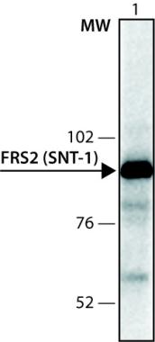
PC-12 cells extract was separated on SDS-PAGE, blotted with 1:2,000 Anti-FRS2 (SNT-1) and developed using Goat Anti-Rabbit IgG-Peroxidase and a chemiluminescent substrate| Product Name | FRS2 antibody |
|---|---|
| Antibody Type | Primary Antibodies |
| Product description | Rabbit polyclonal to FRS2FGFs (Fibroblast Growth Factors) bind to FGF receptors (FGFRs) monovalently, and FGF receptor dimerization and activation is mediated by multivalent interactions between heparin sulfate proteoglycans and FGF. Upon activation, receptor tyrosine kinases undergo rapid autophosphorylation on numerous tyrosine residues. Autophosphorylation sites located within the catalytic domain are crucial for stimulation of kinase activity, while autophosphorylation sites located in other regions are usually involved in the recruitment of cellular target proteins. Fibroblast growth factor or nerve growth factor (NGF) stimulation leads to tyrosine phosphorylation of the FGF Receptor Substrate 2 (FRS2) docking proteins. FRS2 is also designated SNT (Suc1 associated neurotrophic factor target protein). Two structurally similar forms of FRS2 are: FRS2alpha (SNT1) and FRS2 beta(SNT2, FRS3). FRS functions as a lipid-anchored docking protein that is tyrosine phosphorylated and recruited to FGFR upon FGF stimulation. FRS2 plays an important role in linking FGF and NGF with the Ras/MAPK signaling pathway, thus relaying information from the cell surface to the nucleus. FRS2 contains both a consensus myristylation sequence, involved in its recruitment to the cell membrane and a putative phosphotyrosine binding (PTB) domain in its amino-terminus. The interaction of FRS2 with FGFR1 occurs via the PTB domain of FRS2 binding to a 12-residue segment in the juxtamembrane region of FGFR1. FRS2 alpha contains four binding sites for the adaptor protein Grb2 and two binding sites for the protein tyrosine phosphatase Shp2. FGF stimulation leads to phosphorylation of Shp2 on a tyrosine residue forming a complex with an additional molecule of Grb2. Grb2/Sos complexes are thus recruited directly and indirectly via Shp2 upon tyrosine phosphorylation of FRS2alpha in response to growth factor stimulation. It has been shown that FRS2 proteins are tyrosine phosphorylated in response to activation of the RET receptor, a tyrosine kinase that functions as the signal transducing receptor for the GDNF. |
| Immunogen | Synthetic peptide conjugated to KLH, corresponding to amino acids 487-505 of Human FRS2alpha. |
| Clonality | Polyclonal |
|---|---|
| Isotype | IgG |
| Host Species | Rabbit |
| Tested Applications | IPWB |
| For IP: Use 10μg to 20ug using 3 mg RIPA lysate of mouse NIH/3T3 cells. For WB: Use at a dilution of 1/1000. Detects a band of approximately 92 and 95 kDa. Optimal dilutions/concentrations should be determined by the end user.: |
|
| Species Reactivity | HumanMouseRat |
| Concentration | 1mg/ml |
| Purification | Affinity purified |
| Gene Synonyms | FRS2A SNT-1 SNT FRS2 607743 SNT1 10818 Q8WU20 FRS2alpha SNT 1 |
|---|---|
| Target | FRS2 |
Application
PC-12 cells extract was separated on SDS-PAGE, blotted with 1:2,000 Anti-FRS2 (SNT-1) and developed using Goat Anti-Rabbit IgG-Peroxidase and a chemiluminescent substrate| Positive Control | Rat PC-12 and human HepG2 cell lysates |
|---|---|
| Application Notes | For IP: Use 10μg to 20ug using 3 mg RIPA lysate of mouse NIH/3T3 cells. For WB: Use at a dilution of 1/1000. Detects a band of approximately 92 and 95 kDa. Optimal dilutions/concentrations should be determined by the end user.: |
| Form | Liquid |
|---|---|
| Storage Instructions | Keep as concentrated solution. Store at 4ºC short term. For extended storage aliquot and store at -20ºC or below. Avoid freeze-thaw cycles. |
| Storage Buffer | 0.01M PBS, pH 7.4 containing 1% BSA and 15mM Sodium Azide |


