Application
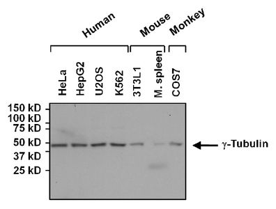
Western blot analysis of gamma Tubulin in 50ug of various whole cell lysates. Proteins were transferred to a PVDF membrane and blocked with 5% Milk/TBST for at least 1 hour. Membranes were probed with gamma Tubulin antibody [4D11] at a dilution of 1:1000 overnight at 4°C on a rocking platform. Membranes were washed in TBS-0.1%Tween 20 and probed with a proper secondary antibody. Membranes were washed and chemiluminescent detection was performed.Application
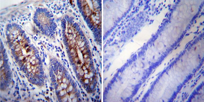
Immunohistochemistry was performed on normal deparaffinized human colon tissue. To expose target proteins, heat induced antigen retrieval was performed using 10mM sodium citrate (pH6.0) buffer, microwaved for 8-15 minutes. Following antigen retrieval tissues were blocked in 3% BSA-PBS for 30 minutes at room temperature. Tissues were then probed at a dilution of 1:200 with or without gamma Tubulin antibody [4D11] overnight at 4°C in a humidified chamber. Tissues were washed extensively with PBST and endogenous peroxidase activity was quenched with a peroxidase suppressor. Detection was performed using a biotin-conjugated secondary antibody and SA-HRP, followed by colorimetric detection using DAB. Tissues were counterstained with hematoxylin and prepped for mounting.Application
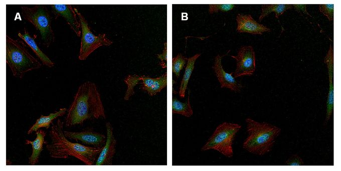
Immunofluorescent analysis of gamma Tubulin (green) in (A) HeLa and (B) U2-OS cells. Formalin-fixed cells were permeabilized with 0.1% Triton X-100 in TBS for 10 minutes at room temperature. Cells were then blocked with 1% BSA for 15 minutes at room temperature. Cells were probed with gamma Tubulin antibody [4D11] at a dilution of 1:50 for at least 1 hour at room temperature. Cells were washed with PBS and incubated with a proper secondary antibody. F-Actin (red) was stained with phalloidin, nuclei (blue) were stained with Hoechst 33342 dye. Images were taken at 20X magnification.| Product Name | gamma Tubulin antibody [4D11] |
|---|---|
| Antibody Type | Primary Antibodies |
| Product description | Mouse monoclonal antibody [4D11] to gamma TubulinThis gene encodes a member of the tubulin superfamily. The encoded protein localizes to the centrosome where it binds to microtubules as part of a complex referred to as the gamma-tubulin ring complex. The protein mediates microtubule nucleation and is required for microtubule formation and progression of the cell cycle. A pseudogene of this gene is found on chromosome 7. [provided by RefSeq, Jan 2009] |
| Immunogen | Recombinant protein containing residues A A T R P D Y I S W G T Q E Q of human gamma Tubulin. |
| Clonality | Monoclonal |
|---|---|
| Isotype | IgG2 |
| Clone Number | 4D11 |
| Host Species | Mouse |
| Tested Applications | ICC/IFIHCIHC-PIPWB |
| ICC/IF: 1:50 | |
| Species Reactivity | HumanMouseRatPrimate |
| Concentration | 1mg/ml (Please refer to the vial label for the specific concentration) |
| Purification | Protein G |
| Gene Synonyms | tubulin, gamma 1 , TUBG1 , GCP-1 , TUBG , TUBGCP1 |
|---|---|
| Target | gamma Tubulin |
Application
Western blot analysis of gamma Tubulin in 50ug of various whole cell lysates. Proteins were transferred to a PVDF membrane and blocked with 5% Milk/TBST for at least 1 hour. Membranes were probed with gamma Tubulin antibody [4D11] at a dilution of 1:1000 overnight at 4°C on a rocking platform. Membranes were washed in TBS-0.1%Tween 20 and probed with a proper secondary antibody. Membranes were washed and chemiluminescent detection was performed.
Application
Immunohistochemistry was performed on normal deparaffinized human colon tissue. To expose target proteins, heat induced antigen retrieval was performed using 10mM sodium citrate (pH6.0) buffer, microwaved for 8-15 minutes. Following antigen retrieval tissues were blocked in 3% BSA-PBS for 30 minutes at room temperature. Tissues were then probed at a dilution of 1:200 with or without gamma Tubulin antibody [4D11] overnight at 4°C in a humidified chamber. Tissues were washed extensively with PBST and endogenous peroxidase activity was quenched with a peroxidase suppressor. Detection was performed using a biotin-conjugated secondary antibody and SA-HRP, followed by colorimetric detection using DAB. Tissues were counterstained with hematoxylin and prepped for mounting.
Application
Immunofluorescent analysis of gamma Tubulin (green) in (A) HeLa and (B) U2-OS cells. Formalin-fixed cells were permeabilized with 0.1% Triton X-100 in TBS for 10 minutes at room temperature. Cells were then blocked with 1% BSA for 15 minutes at room temperature. Cells were probed with gamma Tubulin antibody [4D11] at a dilution of 1:50 for at least 1 hour at room temperature. Cells were washed with PBS and incubated with a proper secondary antibody. F-Actin (red) was stained with phalloidin, nuclei (blue) were stained with Hoechst 33342 dye. Images were taken at 20X magnification.| Application Notes | ICC/IF: 1:50 |
|---|
| Form | Liquid |
|---|---|
| Storage Instructions | -20° C, Avoid Freeze/Thaw Cycles |
| Storage Buffer | PBS containing 1 mg/ml BSA and 0.05 % sodium azide |


