Application
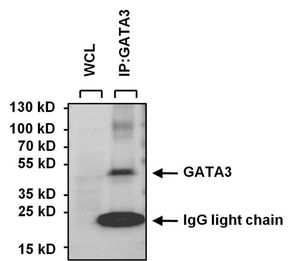
IP analysis was performed with 250ug of SH-SY5Y whole cell lysate and 5ug of GATA3 antibody [1A12-1D9]. The precipitate and 25ug of SH-SY5Y whole cell lysate (loading control) were detected by the same antibody at a dilution of 1:1000.Application
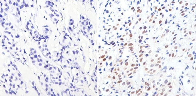
IHC-P analysis of human breast carcinoma with (right) or without (left) GATA3 antibody [1A12-1D9] at a dilution of 1:1000. To expose target proteins, antigen retrieval was performed using 10mM sodium citrate (pH 6.0), microwaved for 8-15 min.Application
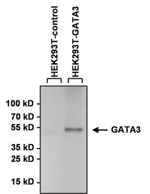
WB analysis of lysates from HEK293T cells overexpressing GATA3-DDK (right) or empty vector control (left) using GATA3 antibody [1A12-1D9] at a dilution of 1:1000.Application
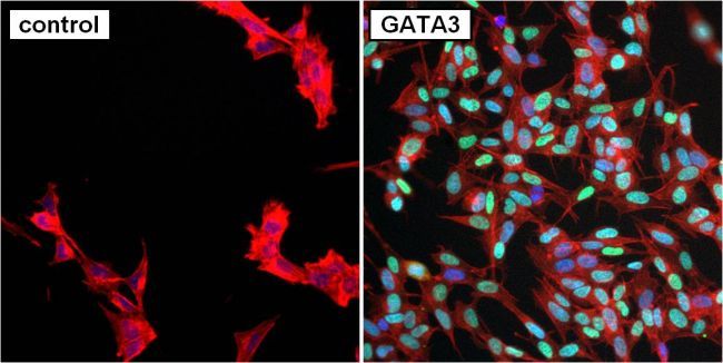
ICC/IF analysis of SH-SY5Y cells without (left) or with (right) GATA3 antibody [1A12-1D9] at a dilution of 1:50 (green). F-Actin (red) was stained with Phalloidin and nuclei (blue) were stained with Hoechst 33342.Application
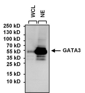
WB analysis of SH-SY5Y whole cell lysate (WCL) and SH-SY5Y nuclear extract (NE) (15ug per lane) using GATA3 antibody [1A12-1D9] at a dilution of 1:1000.Application
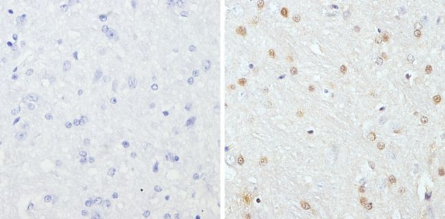
IHC-P analysis of rat brain tissues with (right) or without GATA3 antibody [1A12-1D9] at a dilution of 1:200. To expose target proteins, antigen retrieval was performed using 10mM sodium citrate (pH 6.0), microwaved for 8-15 min.Application
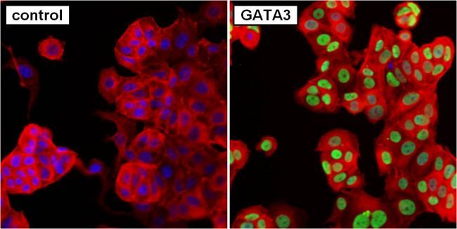
ICC/IF analysis of MCF7 cells without (left) or with (right) GATA3 antibody [1A12-1D9] at a dilution of 1:50 (green). F-Actin (red) was stained with Phalloidin and nuclei (blue) were stained with Hoechst 33342.Application
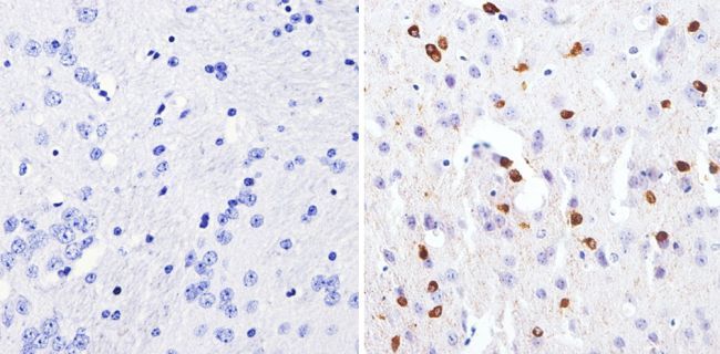
IHC-P analysis of mouse brain tissues with (right) or without (left) GATA3 antibody [1A12-1D9] at a dilution of 1:1000. To expose target proteins, antigen retrieval was performed using 10mM sodium citrate (pH 6.0), microwaved for 8-15 min.| Product Name | GATA3 antibody [1A12-1D9] |
|---|---|
| Antibody Type | Primary Antibodies |
| Product description | Mouse Monoclonal antibody [1A12-1D9] to GATA3This gene encodes a protein which belongs to the GATA family of transcription factors. The protein contains two GATA-type zinc fingers and is an important regulator of T-cell development and plays an important role in endothelial cell biology. Defects in this gene are the cause of hypoparathyroidism with sensorineural deafness and renal dysplasia. [provided by RefSeq, Nov 2009] |
| Immunogen | Purified partial recombinant human GATA3 protein expressed in E. coli. |
| Clonality | Monoclonal |
|---|---|
| Isotype | IgG1 |
| Clone Number | 1A12-1D9 |
| Host Species | Mouse |
| Tested Applications | ICC/IFIHC-PIPWB |
| ICC/IF: 1:50 - 1:100. IHC-P: 1:200 - 1: 2000. IP: 5ug. WB: 1:1000. IHC-P: 1:200 - 1: 2000 | |
| Species Reactivity | Human |
| Concentration | 1mg/ml (Please refer to the vial label for the specific concentration) |
| Purification | Protein A |
| Gene Synonyms | HDRS , GATA binding protein 3 , HDR |
|---|---|
| Target | GATA3 |
Application
IP analysis was performed with 250ug of SH-SY5Y whole cell lysate and 5ug of GATA3 antibody [1A12-1D9]. The precipitate and 25ug of SH-SY5Y whole cell lysate (loading control) were detected by the same antibody at a dilution of 1:1000.
Application
IHC-P analysis of human breast carcinoma with (right) or without (left) GATA3 antibody [1A12-1D9] at a dilution of 1:1000. To expose target proteins, antigen retrieval was performed using 10mM sodium citrate (pH 6.0), microwaved for 8-15 min.
Application
WB analysis of lysates from HEK293T cells overexpressing GATA3-DDK (right) or empty vector control (left) using GATA3 antibody [1A12-1D9] at a dilution of 1:1000.
Application
ICC/IF analysis of SH-SY5Y cells without (left) or with (right) GATA3 antibody [1A12-1D9] at a dilution of 1:50 (green). F-Actin (red) was stained with Phalloidin and nuclei (blue) were stained with Hoechst 33342.
Application
WB analysis of SH-SY5Y whole cell lysate (WCL) and SH-SY5Y nuclear extract (NE) (15ug per lane) using GATA3 antibody [1A12-1D9] at a dilution of 1:1000.
Application
IHC-P analysis of rat brain tissues with (right) or without GATA3 antibody [1A12-1D9] at a dilution of 1:200. To expose target proteins, antigen retrieval was performed using 10mM sodium citrate (pH 6.0), microwaved for 8-15 min.
Application
ICC/IF analysis of MCF7 cells without (left) or with (right) GATA3 antibody [1A12-1D9] at a dilution of 1:50 (green). F-Actin (red) was stained with Phalloidin and nuclei (blue) were stained with Hoechst 33342.
Application
IHC-P analysis of mouse brain tissues with (right) or without (left) GATA3 antibody [1A12-1D9] at a dilution of 1:1000. To expose target proteins, antigen retrieval was performed using 10mM sodium citrate (pH 6.0), microwaved for 8-15 min.| Application Notes | ICC/IF: 1:50 - 1:100. IHC-P: 1:200 - 1: 2000. IP: 5ug. WB: 1:1000. IHC-P: 1:200 - 1: 2000 |
|---|
| Form | Liquid |
|---|---|
| Storage Instructions | -20°C |
| Storage Buffer | PBS with 1mg/ml BSA, 30% glycerol and 0.05% sodium azide |


