Application
HEK293T cells transfected with either pCMV6-ENTRY GBE1Application
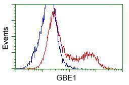
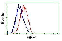
Flow cytometric analysis of Jurkat cells, using anti-GBE1 antibody,(Red) compared to a nonspecific negative control antibody(TA50011)(Blue).Application
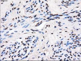
Immunohistochemical staining of paraffin-embedded endometrium tissue using anti-GBE1mouse monoclonal antibody. ( Dilution 1:50)Application
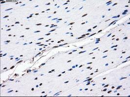
Immunohistochemical staining of paraffin-embedded colon tissue using anti-GBE1mouse monoclonal antibody. ( Dilution 1:50)Application
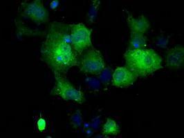
Anti-GBE1 mouse monoclonal antibody immunofluorescent staining of COS7 cells transiently transfected with GBE1Application
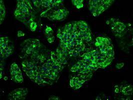
Immunofluorescent staining of HepG2 cells using anti-GBE1 mouse monoclonal antibody .Application
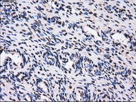
Immunohistochemical staining of paraffin-embedded Ovary tissue using anti-GBE1mouse monoclonal antibody. ( Dilution 1:50)Application
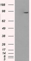
HEK293T cells were transfected with the pCMV6-ENTRY control (Left lane) or pCMV6-ENTRY GBE1 (Right lane) cDNA for 48 hrs and lysed. Equivalent amounts of cell lysates (5 ug per lane) were separated by SDS-PAGE and immunoblotted with anti-GBE1.Application
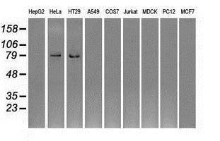
Western blot analysis of extracts (35ug) from 9 different cell lines by using anti-GBE1 monoclonal antibody.| Product Name | GBE1 antibody [1D11] |
|---|---|
| Antibody Type | Primary Antibodies |
| Product description | Mouse monoclonal antibody [1D11] to GBE1Required for sufficient glycogen accumulation. The alpha 1-6 branches of glycogen play an important role in increasing the solubility of the molecule and, consequently, in reducing the osmotic pressure within cells. |
| Immunogen | Full length human recombinant protein of human GBE1 (NP_000149) produced in HEK293T cell. |
| Clonality | Monoclonal |
|---|---|
| Isotype | IgG1 |
| Clone Number | 1D11 |
| Host Species | Mouse |
| Tested Applications | FACSICC/IFIHCIHC-PWB |
| IHC 1:50 | |
| Species Reactivity | Human |
| Concentration | 1mg/ml |
| Purification | Affinity purified |
| Gene Synonyms | GBE |
|---|---|
| Target | GBE1 |
| Tissue Specificity | is specific for human GBE1 (NP_000149). |
Application
HEK293T cells transfected with either pCMV6-ENTRY GBE1
Application
Flow cytometric analysis of Jurkat cells, using anti-GBE1 antibody,(Red) compared to a nonspecific negative control antibody(TA50011)(Blue).
Application
Immunohistochemical staining of paraffin-embedded endometrium tissue using anti-GBE1mouse monoclonal antibody. ( Dilution 1:50)
Application
Immunohistochemical staining of paraffin-embedded colon tissue using anti-GBE1mouse monoclonal antibody. ( Dilution 1:50)
Application
Anti-GBE1 mouse monoclonal antibody immunofluorescent staining of COS7 cells transiently transfected with GBE1
Application
Immunofluorescent staining of HepG2 cells using anti-GBE1 mouse monoclonal antibody .
Application
Immunohistochemical staining of paraffin-embedded Ovary tissue using anti-GBE1mouse monoclonal antibody. ( Dilution 1:50)
Application
HEK293T cells were transfected with the pCMV6-ENTRY control (Left lane) or pCMV6-ENTRY GBE1 (Right lane) cDNA for 48 hrs and lysed. Equivalent amounts of cell lysates (5 ug per lane) were separated by SDS-PAGE and immunoblotted with anti-GBE1.
Application
Western blot analysis of extracts (35ug) from 9 different cell lines by using anti-GBE1 monoclonal antibody.| Application Notes | IHC 1:50 |
|---|
| Form | Liquid |
|---|---|
| Storage Instructions | For short-term storage, store at 4°C or aliquot into working amounts and store at -20°C. For long-term storage, store at -70°C (aliquotted). Avoid repeated freeze-thaw cycles. |
| Storage Buffer | PBS (PH 7.3) containing 1% BSA, 50% glycerol and 0.02% sodium azide. |


