Application
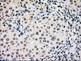
Immunohistochemical staining of paraffin-embedded Adenocarcinoma of breast tissue using anti-GCK mouse monoclonal antibody. ( Dilution 1:50)Application
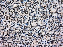
Immunohistochemical staining of paraffin-embedded Carcinoma of thyroid tissue using anti-GCKmouse monoclonal antibody. ( Dilution 1:50)Application
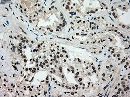
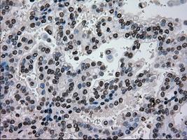
Immunohistochemical staining of paraffin-embedded Carcinoma of prostate tissue using anti-GCKmouse monoclonal antibody. ( Dilution 1:50)Application
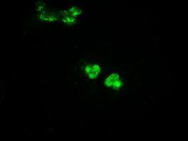
Anti-GCK mouse monoclonal antibody immunofluorescent staining of COS7 cells transiently transfected with GCKApplication
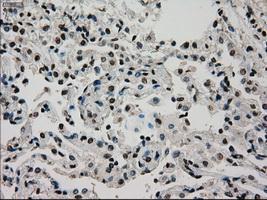
Immunohistochemical staining of paraffin-embedded Carcinoma of lung tissue using anti-GCKmouse monoclonal antibody. ( Dilution 1:50)Application
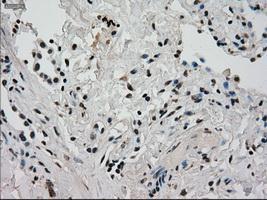
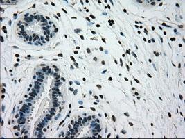
Immunohistochemical staining of paraffin-embedded breast tissue using anti-GCK mouse monoclonal antibody. ( Dilution 1:50)Application
Immunohistochemical staining of paraffin-embedded Carcinoma of kidney tissue using anti-GCKmouse monoclonal antibody. ( Dilution 1:50)Application
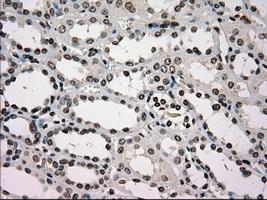
Immunohistochemical staining of paraffin-embedded Kidney tissue using anti-GCKmouse monoclonal antibody. ( Dilution 1:50)Application
Immunohistochemical staining of paraffin-embedded lung tissue using anti-GCKmouse monoclonal antibody. ( Dilution 1:50)Application
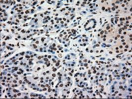
Immunohistochemical staining of paraffin-embedded pancreas tissue using anti-GCKmouse monoclonal antibody. ( Dilution 1:50)Application
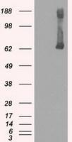
HEK293T cells were transfected with the pCMV6-ENTRY control (Left lane) or pCMV6-ENTRY GCK (Right lane) cDNA for 48 hrs and lysed. Equivalent amounts of cell lysates (5 ug per lane) were separated by SDS-PAGE and immunoblotted with anti-GCK.| Product Name | GCK antibody [3E3] |
|---|---|
| Antibody Type | Primary Antibodies |
| Product description | Mouse monoclonal antibody [3E3] to GCKCatalyzes the initial step in utilization of glucose by the beta-cell and liver at physiological glucose concentration. Glucokinase has a high Km for glucose, and so it is effective only when glucose is abundant. The role of GCK is to provide G6P for the synthesis of glycogen. Pancreatic glucokinase plays an important role in modulating insulin secretion. Hepatic glucokinase helps to facilitate the uptake and conversion of glucose by acting as an insulin-sensitive determinant of hepatic glucose usage. |
| Immunogen | Protein expressed in 293T cell transfected with human GCK expression vector |
| Clonality | Monoclonal |
|---|---|
| Isotype | IgG1 |
| Clone Number | 3E3 |
| Host Species | Mouse |
| Tested Applications | FACSICC/IFIHCIHC-PWB |
| Optimal working dilutions should be determined experimentally by the end user. | |
| Species Reactivity | Human |
| Concentration | 1mg/ml |
| Purification | Affinity purified |
| Gene Synonyms | GCK, HXKP, HK4, MODY2, HKIV, GK, GLK |
|---|---|
| Target | GCK |
| Tissue Specificity | is specific for human GCK. |
Application
Immunohistochemical staining of paraffin-embedded Adenocarcinoma of breast tissue using anti-GCK mouse monoclonal antibody. ( Dilution 1:50)
Application
Immunohistochemical staining of paraffin-embedded Carcinoma of thyroid tissue using anti-GCKmouse monoclonal antibody. ( Dilution 1:50)
Application
Immunohistochemical staining of paraffin-embedded Carcinoma of prostate tissue using anti-GCKmouse monoclonal antibody. ( Dilution 1:50)
Application
Anti-GCK mouse monoclonal antibody immunofluorescent staining of COS7 cells transiently transfected with GCK
Application
Immunohistochemical staining of paraffin-embedded Carcinoma of lung tissue using anti-GCKmouse monoclonal antibody. ( Dilution 1:50)
Application
Immunohistochemical staining of paraffin-embedded breast tissue using anti-GCK mouse monoclonal antibody. ( Dilution 1:50)
Application
Immunohistochemical staining of paraffin-embedded Carcinoma of kidney tissue using anti-GCKmouse monoclonal antibody. ( Dilution 1:50)
Application
Immunohistochemical staining of paraffin-embedded Kidney tissue using anti-GCKmouse monoclonal antibody. ( Dilution 1:50)
Application
Immunohistochemical staining of paraffin-embedded lung tissue using anti-GCKmouse monoclonal antibody. ( Dilution 1:50)
Application
Immunohistochemical staining of paraffin-embedded pancreas tissue using anti-GCKmouse monoclonal antibody. ( Dilution 1:50)
Application
HEK293T cells were transfected with the pCMV6-ENTRY control (Left lane) or pCMV6-ENTRY GCK (Right lane) cDNA for 48 hrs and lysed. Equivalent amounts of cell lysates (5 ug per lane) were separated by SDS-PAGE and immunoblotted with anti-GCK.| Application Notes | Optimal working dilutions should be determined experimentally by the end user. |
|---|
| Form | Liquid |
|---|---|
| Storage Instructions | For short-term storage, store at 4°C or aliquot into working amounts and store at -20°C. For long-term storage, store at -70°C (aliquotted). Avoid repeated freeze-thaw cycles. |
| Storage Buffer | PBS (PH 7.3) containing 1% BSA, 50% glycerol and 0.02% sodium azide. |


