Application
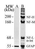
Cytoskeletal homogenate of rat spinal cord was separated by 7.5% SDS-PAGE and stained with Coomassie Brilliant Blue or transferred to nitrocellulose and western blotted with a GFAP antibody. Lane A: Western blot analysis of GFAP with Lane B: SDS| Product Name | GFAP antibody |
|---|---|
| Antibody Type | Primary Antibodies |
| Product description | Mouse monoclonal [2A5] to GFAP - Astrocyte MarkerGlial fibrillary acidic protein (GFAP) is a member of the class III intermediate filament protein family. It is heavily, and specifically, expressed in astrocytes and certain other astroglia in the central nervous system, in satellite cells in peripheral ganglia, and in non-myelinating Schwann cells in peripheral nerves. In addition, neural stem cells frequently strongly express GFAP. Antibodies to GFAP are therefore very useful as markers of astrocytic cells. In addition many types of brain tumor, presumably derived from astrocytic cells, heavily express GFAP. GFAP is also found in the lens epithelium, Kupffer cells of the liver, in some cells in salivary tumors and has been reported in erythrocytes.GFAP runs on SDS-PAGE gels at ~55 kDa, usually associated with lower molecule weight bands which are thought to be proteolytic fragments and alternate transcripts from the single gene. Antibodies to GFAP are useful markers of astrocytic cells and neural stem cells. In addition many types of brain tumors, presumably derived from astrocytic cells, heavily express GFAP. Finally, Alexander's disease was recently shown to be caused by point mutations in protein coding region of the GFAP gene. All forms of Alexander disease are characterized by the presence of Rosenthal fibers, which are GFAP containing cytoplasmic inclusions found in astrocytes. |
| Immunogen | Purified pig spinal cord GFAP. |
| Clonality | Monoclonal |
|---|---|
| Isotype | IgG1 |
| Clone Number | 2A5 |
| Host Species | Mouse |
| Tested Applications | ICC/IFWB |
| Western Blot: 1:500-1:2500. Immunocytochemistry/Immunofluorescence: 1:50-1:150.: |
|
| Species Reactivity | Mammalian speciesAvian |
| Concentration | 1mg/ml |
| Gene Synonyms | P14136 396562 137780 FLJ45472 2670 GFAP |
|---|---|
| Target | GFAP |
| Light Chain Type | kappa |
Application
Cytoskeletal homogenate of rat spinal cord was separated by 7.5% SDS-PAGE and stained with Coomassie Brilliant Blue or transferred to nitrocellulose and western blotted with a GFAP antibody. Lane A: Western blot analysis of GFAP with Lane B: SDS| Application Notes | Western Blot: 1:500-1:2500. Immunocytochemistry/Immunofluorescence: 1:50-1:150.: |
|---|
| Form | Liquid |
|---|---|
| Storage Instructions | Store at -20C. Avoid freeze-thaw cycles. |
| Storage Buffer | 50% glycerol, 1.0 mg/ml BSA and 0.01% Sodium Azide |


