Application
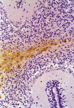
Immunohistochemistry staining of human cerebellum (paraffin-embedded sections) with anti-GFAP (clone GF-01) .Application
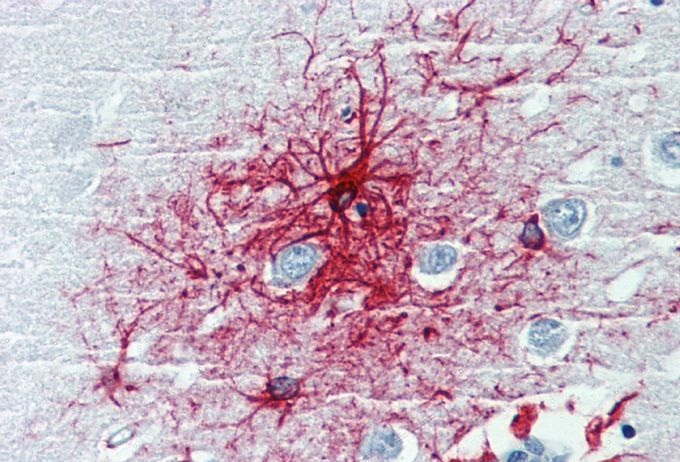
IHC-P analysis of human brain cortex using GFAP antibody [GF-01].| Product Name | GFAP antibody |
|---|---|
| Antibody Type | Primary Antibodies |
| Product description | Mouse monoclonal [GF - 01] to GFAP - Astrocyte MarkerGlial fibrillary acidic protein (GFAP) is a member of the class III intermediate filament protein family. It is heavily and specifically expressed in astrocytes and certain other astroglia in the central nervous system, in satellite cells in peripheral ganglia, and in non-myelinating Schwann cells in peripheral nerves. In addition neural stem cells frequently strongly express GFAP. Antibodies to GFAP are therefore very useful as markers of astrocytic cells. In addition many types of brain tumor, presumably derived from astrocytic cells, heavily express GFAP. GFAP is also found in the lens epithelium, Kupffer cells of the liver, in some cells in salivary tumors and has been reported in erythrocytes. |
| Immunogen | A pellet of pig brain cold-stable proteins after depolymerization of microtubules. |
| Clonality | Monoclonal |
|---|---|
| Isotype | IgG1 |
| Clone Number | GF-01 |
| Host Species | Mouse |
| Tested Applications | ICC/IFIHC-FIHC-PIPWB |
| ICC: Use at an assay dependant dilution. IHC-P: Use at an assay dependant concentration. The antibody strongly stains astrocytes in human brain tissue sections but it is essentially negative on mouse and rat tissues. IHC-Fr: Use at an assay dependant concentration. IP: Use at an assay dependant dilution. WB: Use at an assay dependant concentration. Positive in dibutyryl cAMP stimulated human glioma cell line U333 CG/343 MG, but negative in rat glioma cell line C6. Positive in pig brain lysate, but negative in mouse brain lysate. Optimal dilutions/concentrations should be determined by the end user.: |
|
| Species Reactivity | HumanPigCat |
| Concentration | 1mg/ml (Please refer to the vial label for the specific concentration) |
| Purification | Protein A |
| Gene Synonyms | P14136 396562 137780 FLJ45472 2670 GFAP |
|---|---|
| Target | GFAP |
Application
Immunohistochemistry staining of human cerebellum (paraffin-embedded sections) with anti-GFAP (clone GF-01) .
Application
IHC-P analysis of human brain cortex using GFAP antibody [GF-01].| Application Notes | ICC: Use at an assay dependant dilution. IHC-P: Use at an assay dependant concentration. The antibody strongly stains astrocytes in human brain tissue sections but it is essentially negative on mouse and rat tissues. IHC-Fr: Use at an assay dependant concentration. IP: Use at an assay dependant dilution. WB: Use at an assay dependant concentration. Positive in dibutyryl cAMP stimulated human glioma cell line U333 CG/343 MG, but negative in rat glioma cell line C6. Positive in pig brain lysate, but negative in mouse brain lysate. Optimal dilutions/concentrations should be determined by the end user.: |
|---|
| Form | Liquid |
|---|---|
| Storage Instructions | Keep as concentrated solution. Store at 4°C short term. For extended storage aliquot and store at -20°C or below. Avoid freeze-thaw cycles. |
| Storage Buffer | Phosphate buffered saline pH 7.4 containing 15mM sodium azide |


