Application
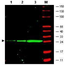
Figure: Western blot of GFP recombinant protein detected with GeneTex monoclonal anti-GFP antibody. GFP recombinant protein was expressed in HeLa cells, where 50 ng (lane 1), 100 ng (lane 2) and 500 ng (lane 3) of lysate were loaded per lane. detects a 27 kDa band corresponding to the epitope tag GFP. The membrane was probed with the primary antibody for 1 h at room temperature followed by washes and reaction with a 1:2,500 dilution of IRDye® 800 conjugated goat anti-Mouse IgG [H&L]. IRDye® 800 fluorescence image was captured using the Odyssey® Infrared Imaging System developed by LI-COR. IRDye is a trademark of LI-COR, Inc. Other detection systems will yield similar results.Application
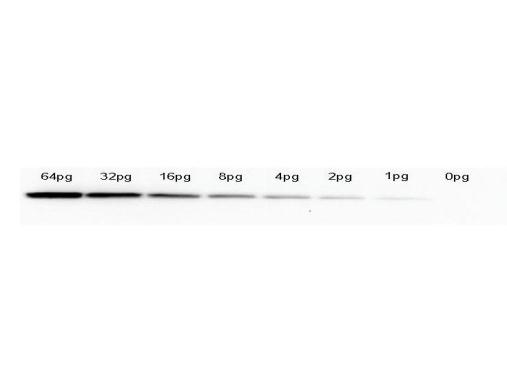
Western Blot of anti-GFP monoclonal antibody . Lane 1: 64pg of recombinant GFP protein were spiked into a HeLa cell-derived lysates). Lane 2: 32pg of recombinant GFP protein were spiked into a HeLa cell-derived lysates. Lane 3: 16pg of recombinant GFP protein were spiked into a HeLa cell-derived lysates. Lane 4: 8pg of recombinant GFP protein were spiked into a HeLa cell-derived lysates. Lane 5: 4pg of recombinant GFP protein were spiked into a HeLa cell-derived lysates. Lane 6: 2pg of recombinant GFP protein were spiked into a HeLa cell-derived lysates. Lane 7: 1g of recombinant GFP protein were spiked into a HeLa cell-derived lysates. Lane 8: 0pg of recombinant GFP protein were spiked into a HeLa cell-derived lysates. Primary antibody: anti-GFP monoclonal antibody at 1:1000 for overnight at 4°C. Secondary antibody: HRP-conjugated anti-Mouse IgG was performed at a dilution of 1:20,000 for 1h at 4°C. Block: TTBS supplemented with 1% BSA for 1 h at 4°C. Predicted/Observed size: 27 kDa for GFP. Other band(s): none.| Product Name | GFP antibody [9F9.F9] |
|---|---|
| Antibody Type | Tags Antibodies |
| Product description | Mouse monoclonal [9F9.F9] antibody to GFPThe jellyfish Aequorea victoria contains green fluorescent protein (GFP) that emits light in the bioluminescence reaction of the animal. GFP has been used widely as a reporter protein for gene expression in eukaryotic and prokaryotic organisms, and as a protein tag in cell culture and in multicellular organisms. As a fusion tag, GFP can be used to localize proteins, to study their movement or to research the dynamics of the subcellular compartments where these proteins are targeted. GFP technology has revealed considerable new insights into the physiological activities of living cells. GFP is a 27 kDa monomeric protein, which autocatalytically forms a fluorescent pigment. The wild type protein absorbs blue light (maximally at 395nm) and emits green light (peak emission 508nm) in the absence of additional proteins, substrates, or co-factors. GFP fluorescence is stable, species independent and is suitable for a variety of applications. GFP has been used extensively as a fluorescent tag to monitor gene expression and protein localization. Moreover, other applications for GFP include its use in assessing protein protein interactions in the yeast two hybrid system, and in measuring distances between proteins in fluorescence energy transfer (FRET) experiments. |
| Immunogen | The immunogen is a Green Fluorescent Protein (GFP) fusion protein corresponding to the full length amino acid sequence (246aa) derived from the jellyfish Aequorea victoria. |
| Clonality | Monoclonal |
|---|---|
| Isotype | IgG2a |
| Clone Number | 9F9.F9 |
| Host Species | Mouse |
| Tested Applications | ELISAICC/IFIHCIPWB |
| For IHC: Use at a dilution of 1:100-1:500. For WB: Use at a dilution of 1:1000-1:4000. Optimal dilutions/concentrations should be determined by the researcher. | |
| Concentration | 1mg/ml |
| Purification | Unpurified |
| Gene Synonyms | eGFP, Green Fluorescent Protein, GFP, wtGFP |
|---|---|
| Target | GFP |
Application
Figure: Western blot of GFP recombinant protein detected with GeneTex monoclonal anti-GFP antibody. GFP recombinant protein was expressed in HeLa cells, where 50 ng (lane 1), 100 ng (lane 2) and 500 ng (lane 3) of lysate were loaded per lane. detects a 27 kDa band corresponding to the epitope tag GFP. The membrane was probed with the primary antibody for 1 h at room temperature followed by washes and reaction with a 1:2,500 dilution of IRDye® 800 conjugated goat anti-Mouse IgG [H&L]. IRDye® 800 fluorescence image was captured using the Odyssey® Infrared Imaging System developed by LI-COR. IRDye is a trademark of LI-COR, Inc. Other detection systems will yield similar results.
Application
Western Blot of anti-GFP monoclonal antibody . Lane 1: 64pg of recombinant GFP protein were spiked into a HeLa cell-derived lysates). Lane 2: 32pg of recombinant GFP protein were spiked into a HeLa cell-derived lysates. Lane 3: 16pg of recombinant GFP protein were spiked into a HeLa cell-derived lysates. Lane 4: 8pg of recombinant GFP protein were spiked into a HeLa cell-derived lysates. Lane 5: 4pg of recombinant GFP protein were spiked into a HeLa cell-derived lysates. Lane 6: 2pg of recombinant GFP protein were spiked into a HeLa cell-derived lysates. Lane 7: 1g of recombinant GFP protein were spiked into a HeLa cell-derived lysates. Lane 8: 0pg of recombinant GFP protein were spiked into a HeLa cell-derived lysates. Primary antibody: anti-GFP monoclonal antibody at 1:1000 for overnight at 4°C. Secondary antibody: HRP-conjugated anti-Mouse IgG was performed at a dilution of 1:20,000 for 1h at 4°C. Block: TTBS supplemented with 1% BSA for 1 h at 4°C. Predicted/Observed size: 27 kDa for GFP. Other band(s): none.| Application Notes | For IHC: Use at a dilution of 1:100-1:500. For WB: Use at a dilution of 1:1000-1:4000. Optimal dilutions/concentrations should be determined by the researcher. |
|---|
| Form | Liquid |
|---|---|
| Storage Instructions | Keep as concentrated solution. Store at 4ºC short term. For extended storage aliquot and store at -20ºC or below. Avoid freeze-thaw cycles. |
| Storage Buffer | 0.02M Potassium phosphate, 0.15M sodium chloride, pH 7.2, containing 0.01% sodium azide |


