Application
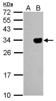
GFP antibody detects GFP protein by western blot analysis. A. 5 µg 293T whole cell extract B. 5 µg 293T whole cell extract expressing GFP-tagged protein 12% SDS-PAGE GFP antibody dilution: 1:5000 The HRP-conjugated anti-mouse IgG antibody was used to detect the primary antibody.Application
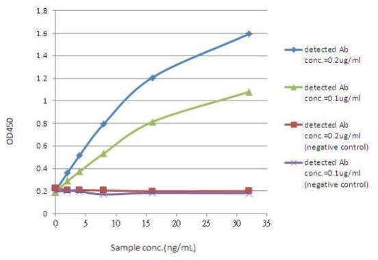
ELISA detection of GFP using for capture and for detection. Coating Ab: mouse anti-GFP (, 2µg/well ) Sample: GFP protein Detected Ab: rabbit anti-GFP Conjugated Ab: goat anti-rabbit IgG-HRP ( 1:20000)Application
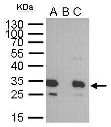
GFP tag antibody immunoprecipitates GFP-fused protein in IP experiments. IP Sample: 400 µg cell lysate/extract of GFP-fused gene transfected 293T cells A. 30 μg lysate/extract of transfected 293T cell B. Control with 2 μg of preimmune mouse IgG C. Immunoprecipitation of GFP-fused protein by 2 μg of GFP tag antibody 12% SDS-PAGE The immunoprecipitated GFP-fused protein was detected by GFP tag antibody diluted at 1:1000. EasyBlot anti-mouse IgG was used as a secondary reagent.Application
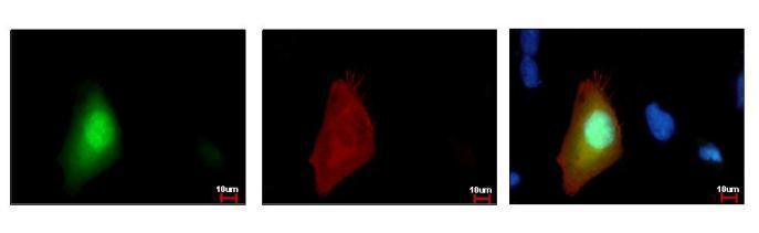
GFP-tag antibody detects GFP-recombinant protein by immunofluorescent analysis. Sample: GFP-tag transfected HeLa cells were fixed in 4% paraformaldehyde at RT for 15 min. Green: GFP is expressed in the tranfected cell Red: .GFP-recombinant protein stained by GFP-tag antibody diluted at 1:200. Blue: Hoechst 33342 staining.| Product Name | GFP antibody [GT859] |
|---|---|
| Antibody Type | Tags Antibodies |
| Product description | Mouse Monoclonal antibody to GFP (Green Fluorescent Protein) |
| Clonality | Monoclonal |
|---|---|
| Isotype | IgG2 |
| Clone Number | GT859 |
| Host Species | Mouse |
| Tested Applications | ELISAICC/IFIHC-FIHC-PIHC-WIPWB |
| WB:1:500~1:2000 IHC:1:50~1:200 IF/ICC:1:100~1:200 *Optimal dilutions/concentrations should be determined by the researcher. |
|
| Species Reactivity | All |
| Concentration | 1mg/ml (Please refer to the vial label for the specific concentration) |
| Purification | Affinity purified |
| Tissue Specificity | This antibody reacts with GFP variants including GFP, EGFP, and YFP. |
|---|
Application
GFP antibody detects GFP protein by western blot analysis. A. 5 µg 293T whole cell extract B. 5 µg 293T whole cell extract expressing GFP-tagged protein 12% SDS-PAGE GFP antibody dilution: 1:5000 The HRP-conjugated anti-mouse IgG antibody was used to detect the primary antibody.
Application
ELISA detection of GFP using for capture and for detection. Coating Ab: mouse anti-GFP (, 2µg/well ) Sample: GFP protein Detected Ab: rabbit anti-GFP Conjugated Ab: goat anti-rabbit IgG-HRP ( 1:20000)
Application
GFP tag antibody immunoprecipitates GFP-fused protein in IP experiments. IP Sample: 400 µg cell lysate/extract of GFP-fused gene transfected 293T cells A. 30 μg lysate/extract of transfected 293T cell B. Control with 2 μg of preimmune mouse IgG C. Immunoprecipitation of GFP-fused protein by 2 μg of GFP tag antibody 12% SDS-PAGE The immunoprecipitated GFP-fused protein was detected by GFP tag antibody diluted at 1:1000. EasyBlot anti-mouse IgG was used as a secondary reagent.
Application
GFP-tag antibody detects GFP-recombinant protein by immunofluorescent analysis. Sample: GFP-tag transfected HeLa cells were fixed in 4% paraformaldehyde at RT for 15 min. Green: GFP is expressed in the tranfected cell Red: .GFP-recombinant protein stained by GFP-tag antibody diluted at 1:200. Blue: Hoechst 33342 staining.| Positive Control | 293T whole cell extract expressing GFP-tagged protein |
|---|---|
| Application Notes | WB:1:500~1:2000 IHC:1:50~1:200 IF/ICC:1:100~1:200 *Optimal dilutions/concentrations should be determined by the researcher. |
| Form | Liquid |
|---|---|
| Storage Instructions | Keep as concentrated solution. Aliquot and store at -20ºC or below. Avoid multiple freeze-thaw cycles. |
| Storage Buffer | PBS |


