Application
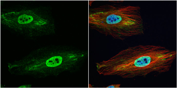
GSTP1 antibody detects GSTP1 protein at cytoplasm and nucleus by immunofluorescent analysis. Sample: HeLa cells were fixed in 4% paraformaldehyde at RT for 15 min. Green: GSTP1 protein stained by GSTP1 antibody diluted at 1:500. Red: alpha Tubulin, a cytoskeleton marker, stained by alpha Tubulin antibody [GT114] diluted at 1:1000. Blue: Hoechst 33342 staining.Application
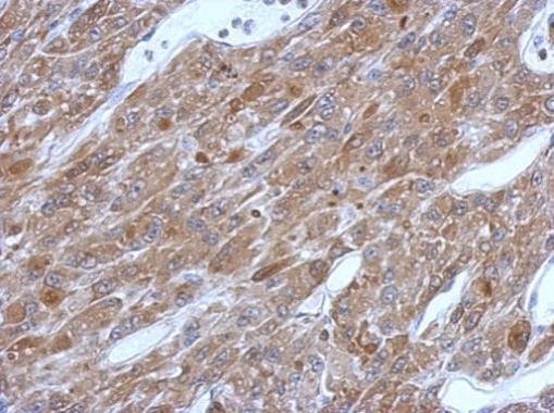
Immunohistochemical analysis of paraffin-embedded U87 xenograft, using GSTP1 antibody at 1:500 dilution.Application
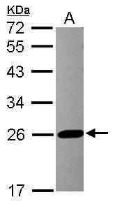
Sample (30 µg of whole cell lysate) A: HCT116 12% SDS PAGE diluted at 1:10000 The HRP-conjugated anti-rabbit IgG antibody was used to detect the primary antibody.Application
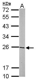
Sample (50 µg of whole cell lysate) A: mouse brain 12% SDS PAGE diluted at 1:1000 The HRP-conjugated anti-rabbit IgG antibody was used to detect the primary antibody.Application
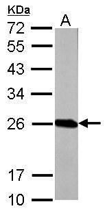
GSTP1 antibody detects GSTP1 protein by western blot analysis. A. 50 µg Rat brain lysate/extract 12% SDS-PAGE GSTP1 antibody dilution: 1:1000 The HRP-conjugated anti-rabbit IgG antibody was used to detect the primary antibody.Application
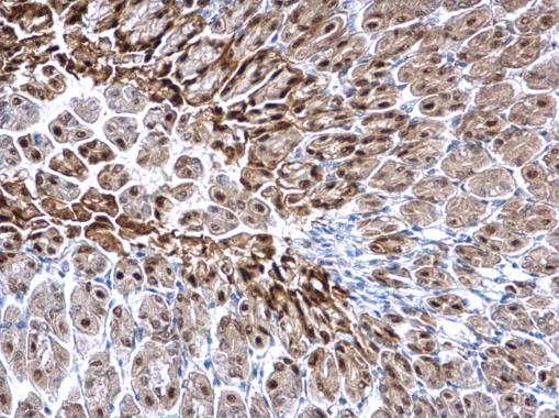
GSTP1 antibody detects GSTP1 protein at nucleus on mouse stomach by immunohistochemical analysis. Sample: Paraffin-embedded mouse stomach. GSTP1 antibody dilution: 1:500.Application
GSTP1 antibody detects GSTP1 protein at nucleus on mouse brain stem by immunohistochemical analysis. Sample: Paraffin-embedded mouse brain stem. GSTP1 antibody dilution: 1:500.Application
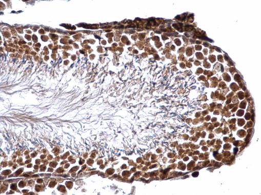
GSTP1 antibody detects GSTP1 protein at nucleus on mouse testis by immunohistochemical analysis. Sample: Paraffin-embedded mouse testis. GSTP1 antibody dilution: 1:500.Application
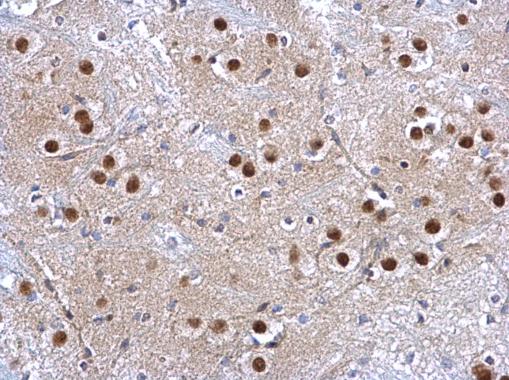
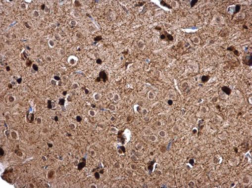
GSTP1 antibody detects GSTP1 protein at nucleus on rat fore brain by immunohistochemical analysis. Sample: Paraffin-embedded rat fore brain. GSTP1 antibody dilution: 1:500.| Product Name | GSTP1 antibody |
|---|---|
| Antibody Type | Primary Antibodies |
| Product description | Rabbit Polyclonal antibody to GSTP1 (glutathione S-transferase pi 1)Glutathione S-transferases (GSTs) are a family of enzymes that play an important role in detoxification by catalyzing the conjugation of many hydrophobic and electrophilic compounds with reduced glutathione. Based on their biochemical, immunologic, and structural properties, the soluble GSTs are categorized into 4 main classes: alpha, mu, pi, and theta. This GST family member is a polymorphic gene encoding active, functionally different GSTP1 variant proteins that are thought to function in xenobiotic metabolism and play a role in susceptibility to cancer, and other diseases. [provided by RefSeq] |
| Immunogen | Recombinant protein encompassing a sequence within the center region of human GSTP1. The exact sequence is proprietary. |
| Clonality | Polyclonal |
|---|---|
| Isotype | IgG |
| Host Species | Rabbit |
| Tested Applications | ICC/IFIHC-PWB |
| *Optimal dilutions/concentrations should be determined by the researcher.
: |
|
| Species Reactivity | HumanMouseRat |
| Predicted Reactivity | Dog, Rhesus Monkey, BovineDog (86%), Rhesus Monkey (97%), Bovine (85%) |
| Concentration | 1mg/ml (Please refer to the vial label for the specific concentration) |
| Purification | Affinity purified |
| Gene Synonyms | DFN7 antibody FAEES3 antibody GST3 antibody GSTP antibody PI antibody GSTP1 antibody deafness X-linked 7 antibody fatty acid ethyl ester synthase III antibody GST class-pi antibody GSTP1-1 antibody glutathione S-transferase P antibody glutathio |
|---|---|
| Molecular Weight(MW) | 23 kDa (note) |
Application
GSTP1 antibody detects GSTP1 protein at cytoplasm and nucleus by immunofluorescent analysis. Sample: HeLa cells were fixed in 4% paraformaldehyde at RT for 15 min. Green: GSTP1 protein stained by GSTP1 antibody diluted at 1:500. Red: alpha Tubulin, a cytoskeleton marker, stained by alpha Tubulin antibody [GT114] diluted at 1:1000. Blue: Hoechst 33342 staining.
Application
Immunohistochemical analysis of paraffin-embedded U87 xenograft, using GSTP1 antibody at 1:500 dilution.
Application
Sample (30 µg of whole cell lysate) A: HCT116 12% SDS PAGE diluted at 1:10000 The HRP-conjugated anti-rabbit IgG antibody was used to detect the primary antibody.
Application
Sample (50 µg of whole cell lysate) A: mouse brain 12% SDS PAGE diluted at 1:1000 The HRP-conjugated anti-rabbit IgG antibody was used to detect the primary antibody.
Application
GSTP1 antibody detects GSTP1 protein by western blot analysis. A. 50 µg Rat brain lysate/extract 12% SDS-PAGE GSTP1 antibody dilution: 1:1000 The HRP-conjugated anti-rabbit IgG antibody was used to detect the primary antibody.
Application
GSTP1 antibody detects GSTP1 protein at nucleus on mouse stomach by immunohistochemical analysis. Sample: Paraffin-embedded mouse stomach. GSTP1 antibody dilution: 1:500.
Application
GSTP1 antibody detects GSTP1 protein at nucleus on mouse brain stem by immunohistochemical analysis. Sample: Paraffin-embedded mouse brain stem. GSTP1 antibody dilution: 1:500.
Application
GSTP1 antibody detects GSTP1 protein at nucleus on mouse testis by immunohistochemical analysis. Sample: Paraffin-embedded mouse testis. GSTP1 antibody dilution: 1:500.
Application
GSTP1 antibody detects GSTP1 protein at nucleus on rat fore brain by immunohistochemical analysis. Sample: Paraffin-embedded rat fore brain. GSTP1 antibody dilution: 1:500.| Positive Control | HCT116 , mouse brain , Rat brain |
|---|---|
| Application Notes | *Optimal dilutions/concentrations should be determined by the researcher.
: |
| Form | Liquid |
|---|---|
| Storage Instructions | Keep as concentrated solution. Aliquot and store at -20ºC or below. Avoid multiple freeze-thaw cycles. |
| Storage Buffer | 0.1M Tris, 0.1M Glycine, 20% Glycerol (pH7). 0.01% Thimerosal was added as a preservative. |


