Application
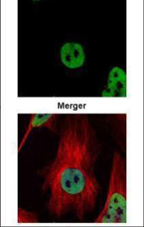
Confocal immunofluorescence analysis (Olympus FV10i) of paraformaldehyde-fixed HeLa, using HDAC2 antibody (Green) at 1:500 dilution. Alpha-tubulin filaments were labeled with (Red) at 1:500.Application
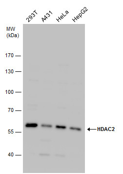
HDAC2 antibody detects HDAC2 protein by western blot analysis. Various whole cell extracts (30 µg) were separated by 7.5% SDS-PAGE, and the membrane was blotted with HDAC2 antibody diluted by 1:1000. The HRP-conjugated anti-rabbit IgG antibody was used to detect the primary antibody.Application
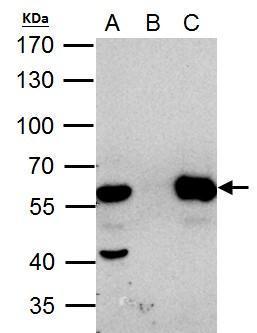
HDAC2 antibody immunoprecipitates HDAC2 protein in IP experiments. IP Sample: HeLa whole cell lysate/extract A. 40 μg HeLa whole cell lysate/extract B. Control with 2 μg of preimmune rabbit IgG C. Immunoprecipitation of HDAC2 protein by 2 μg of HDAC2 antibody 7.5% SDS-PAGE The immunoprecipitated HDAC2 protein was detected by HDAC2 antibody diluted at 1:1000. EasyBlot anti-rabbit IgG was used as a secondary reagent.Application
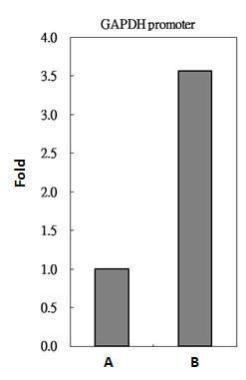
HDAC2 antibody immunoprecipitates HDAC2 protein-DNA complex in ChIP experiments. ChIP Sample: 293T whole cell lysate/extract A. 5 μg preimmune rabbit IgG B. 5 μg of HDAC2 antibody The precipitated DNA was detected by PCR with primer set targeting to GAPDH promoter.Application
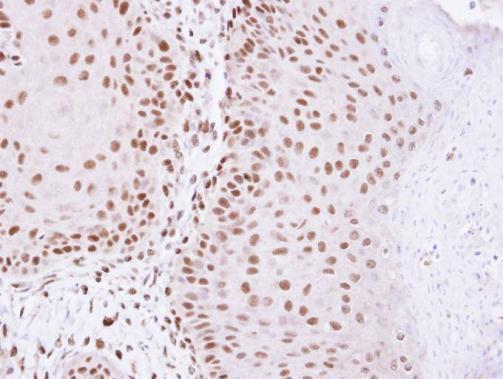
Immunohistochemical analysis of paraffin-embedded Cal27 Xenograft, using HDAC2 antibody at 1:100 dilution.Application
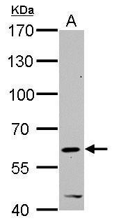
HDAC2 antibody detects HDAC2 protein by western blot analysis. A. 30 µg C2C12 whole cell lysate/extract 7.5% SDS-PAGE HDAC2 antibody dilution: 1:1000 The HRP-conjugated anti-rabbit IgG antibody was used to detect the primary antibody.Application
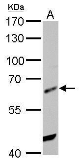
HDAC2 antibody detects HDAC2 protein by western blot analysis. A. 30 µg Rat2 whole cell lysate/extract 7.5% SDS-PAGE HDAC2 antibody dilution: 1:1000 The HRP-conjugated anti-rabbit IgG antibody was used to detect the primary antibody.| Product Name | HDAC2 antibody |
|---|---|
| Antibody Type | Primary Antibodies |
| Product description | Rabbit Polyclonal antibody to HDAC2 (histone deacetylase 2)This gene product belongs to the histone deacetylase family. Histone deacetylases act via the formation of large multiprotein complexes and are responsible for the deacetylation of lysine residues on the N-terminal region of the core histones (H2A, H2B, H3 and H4). This protein also forms transcriptional repressor complexes by associating with many different proteins, including YY1, a mammalian zinc-finger transcription factor. Thus it plays an important role in transcriptional regulation, cell cycle progression and developmental events. [provided by RefSeq] |
| Immunogen | Carrier-protein conjugated synthetic peptide encompassing a sequence within the C-terminus region of human HDAC2. The exact sequence is proprietary. |
| Clonality | Polyclonal |
|---|---|
| Isotype | IgG |
| Host Species | Rabbit |
| Tested Applications | ICC/IFIHC-PIPWBChIP |
| *Optimal dilutions/concentrations should be determined by the researcher. | |
| Species Reactivity | HumanMouseRat |
| Concentration | 1mg/ml (Please refer to the vial label for the specific concentration) |
| Purification | Affinity purified |
| Gene Synonyms | HD2 antibody, RPD3 antibody, YAF1 antibody, HDAC2 antibody, histone deacetylase 2 antibody, transcriptional regulator homolog RPD3 antibody, YY1-associated factor 1 antibody |
|---|---|
| Molecular Weight(MW) | 55 kDa (note) |
| Tissue Specificity | Knockdown/Knockout validation was supported by customer review data. |
| Cellular Localization | Nucleus |
Application
Confocal immunofluorescence analysis (Olympus FV10i) of paraformaldehyde-fixed HeLa, using HDAC2 antibody (Green) at 1:500 dilution. Alpha-tubulin filaments were labeled with (Red) at 1:500.
Application
HDAC2 antibody detects HDAC2 protein by western blot analysis. Various whole cell extracts (30 µg) were separated by 7.5% SDS-PAGE, and the membrane was blotted with HDAC2 antibody diluted by 1:1000. The HRP-conjugated anti-rabbit IgG antibody was used to detect the primary antibody.
Application
HDAC2 antibody immunoprecipitates HDAC2 protein in IP experiments. IP Sample: HeLa whole cell lysate/extract A. 40 μg HeLa whole cell lysate/extract B. Control with 2 μg of preimmune rabbit IgG C. Immunoprecipitation of HDAC2 protein by 2 μg of HDAC2 antibody 7.5% SDS-PAGE The immunoprecipitated HDAC2 protein was detected by HDAC2 antibody diluted at 1:1000. EasyBlot anti-rabbit IgG was used as a secondary reagent.
Application
HDAC2 antibody immunoprecipitates HDAC2 protein-DNA complex in ChIP experiments. ChIP Sample: 293T whole cell lysate/extract A. 5 μg preimmune rabbit IgG B. 5 μg of HDAC2 antibody The precipitated DNA was detected by PCR with primer set targeting to GAPDH promoter.
Application
Immunohistochemical analysis of paraffin-embedded Cal27 Xenograft, using HDAC2 antibody at 1:100 dilution.
Application
HDAC2 antibody detects HDAC2 protein by western blot analysis. A. 30 µg C2C12 whole cell lysate/extract 7.5% SDS-PAGE HDAC2 antibody dilution: 1:1000 The HRP-conjugated anti-rabbit IgG antibody was used to detect the primary antibody.
Application
HDAC2 antibody detects HDAC2 protein by western blot analysis. A. 30 µg Rat2 whole cell lysate/extract 7.5% SDS-PAGE HDAC2 antibody dilution: 1:1000 The HRP-conjugated anti-rabbit IgG antibody was used to detect the primary antibody.| Positive Control | 293T , A431 , HeLa , HepG2 , C2C12 , Rat2 , *NB2a |
|---|---|
| Application Notes | *Optimal dilutions/concentrations should be determined by the researcher. |
| Form | Liquid |
|---|---|
| Storage Instructions | Keep as concentrated solution. Aliquot and store at -20ºC or below. Avoid multiple freeze-thaw cycles. |
| Storage Buffer | 0.1M Tris, 0.1M Glycine, 10% Glycerol (pH7). 0.01% Thimerosal was added as a preservative. |


