Application
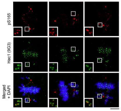
Mouse anti-Hec1 (9G3, cat# 70268) and rabbit anti-Hec1 (phospho Ser165) antibodies were used to co-stain MCF10A cells. Insets show kinetochores of misaligned chromosomes. DAPI staining shows chromatin.Application
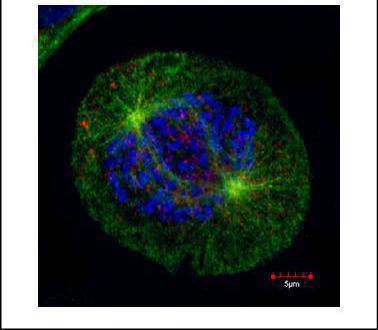
Confocal immunofluorescence staining (Olympus FV10i) of HEC1, a kinetochore outer layer marker. U2OS cells were fixed by 4% PFA and costained with Hec1 9G3.23 AB (Red; cat# 1:500 Ab dilution) and alpha-tubulin rabbit polyclonal AB (Green; cat# 1:500 Ab dilution ), a spindle marker. DAPI (blue), chromosomes. Scale bar, 5 umApplication
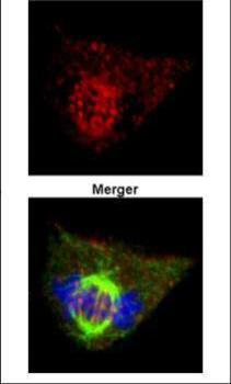
Confocal immunofluorescence analysis (Olympus FV10i) of paraformaldehyde-fixed 293T, using HEC1 antibody (Green) at 1:500 dilution. Alpha-tubulin filaments were labeled with (Red) at 1:500.Application
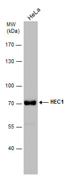
Whole cell extract (30 µg) was separated by 7.5% SDS-PAGE, and the membrane was blotted with HEC1 antibody [9G3.23] diluted at 1:1000. The HRP-conjugated anti-mouse IgG antibody was used to detect the primary antibody, and the signal was developed with Trident ECL plus-Enhanced.Application
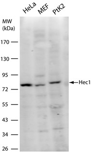
Hec1 [9G3] antibody detects Hec1 [9G3] protein by western blot analysis. Various whole cell extracts (30 µg) were separated by 10% SDS-PAGE, and the membrane was blotted with Hec1 [9G3] antibody diluted at a dilution of 1:1000. The HRP-conjugated anti-mouse IgG antibody was used to detect the primary antibody.Application
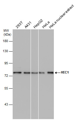
HEC1 antibody [9G3.23] detects HEC1 protein by western blot analysis. Various whole cell extracts and HeLa nuclear extracts (30 µg) were separated by 7.5% SDS-PAGE, and the membrane was blotted with HEC1 antibody [9G3.23] diluted at 1:1000. The HRP-conjugated anti-mouse IgG antibody was used to detect the primary antibody, and the signal was developed with Trident femto Western HRP Substrate .| Product Name | HEC1 antibody |
|---|---|
| Antibody Type | Primary Antibodies |
| Product description | Mouse monoclonal antibody to human HEC1Human HEC 1 is a coiled-coil protein that has been demonstrated to have a role in M-phase progression. It is essential for proper chromosome segregation. Its depletion results inthe impairment of chromosome congression and results in spindle checkpoint activation. |
| Immunogen | Human HEC1 protein consisting of amino acids 56-642. |
| Clonality | Monoclonal |
|---|---|
| Isotype | IgG2a |
| Clone Number | 9G3.23 |
| Host Species | Mouse |
| Tested Applications | FACSICC/IFIHCIPWB |
| For ICC/IF: Use at a concentration of 4-10 µg/ml. For IP: Use at a concentration of 1-5 µg/ml. For WB: Use at a dilution of 1:500-1:3000.For FACS:Assay dependent Optimal dilutions/concentrations should be determined by the researcher.: |
|
| Species Reactivity | HumanMousePigRatHamsterKangarooAfrican Green Monkey |
| Concentration | 0.2mg/ml |
| Purification | Affinity purified |
| Gene Synonyms | HEC 10403 607272 O14777 KNTC2 HEC1 |
|---|---|
| Target | HEC1 |
| Molecular Weight(MW) | 80 kDa (note) |
| Cellular Localization | Nuclear |
Application
Mouse anti-Hec1 (9G3, cat# 70268) and rabbit anti-Hec1 (phospho Ser165) antibodies were used to co-stain MCF10A cells. Insets show kinetochores of misaligned chromosomes. DAPI staining shows chromatin.
Application
Confocal immunofluorescence staining (Olympus FV10i) of HEC1, a kinetochore outer layer marker. U2OS cells were fixed by 4% PFA and costained with Hec1 9G3.23 AB (Red; cat# 1:500 Ab dilution) and alpha-tubulin rabbit polyclonal AB (Green; cat# 1:500 Ab dilution ), a spindle marker. DAPI (blue), chromosomes. Scale bar, 5 um
Application
Confocal immunofluorescence analysis (Olympus FV10i) of paraformaldehyde-fixed 293T, using HEC1 antibody (Green) at 1:500 dilution. Alpha-tubulin filaments were labeled with (Red) at 1:500.
Application
Whole cell extract (30 µg) was separated by 7.5% SDS-PAGE, and the membrane was blotted with HEC1 antibody [9G3.23] diluted at 1:1000. The HRP-conjugated anti-mouse IgG antibody was used to detect the primary antibody, and the signal was developed with Trident ECL plus-Enhanced.
Application
Hec1 [9G3] antibody detects Hec1 [9G3] protein by western blot analysis. Various whole cell extracts (30 µg) were separated by 10% SDS-PAGE, and the membrane was blotted with Hec1 [9G3] antibody diluted at a dilution of 1:1000. The HRP-conjugated anti-mouse IgG antibody was used to detect the primary antibody.
Application
HEC1 antibody [9G3.23] detects HEC1 protein by western blot analysis. Various whole cell extracts and HeLa nuclear extracts (30 µg) were separated by 7.5% SDS-PAGE, and the membrane was blotted with HEC1 antibody [9G3.23] diluted at 1:1000. The HRP-conjugated anti-mouse IgG antibody was used to detect the primary antibody, and the signal was developed with Trident femto Western HRP Substrate .| Positive Control | HeLa , MEF , PtK2 , 293T , A431 , HepG2 , U87-MG NE nuclear extract |
|---|---|
| Application Notes | For ICC/IF: Use at a concentration of 4-10 µg/ml. For IP: Use at a concentration of 1-5 µg/ml. For WB: Use at a dilution of 1:500-1:3000.For FACS:Assay dependent Optimal dilutions/concentrations should be determined by the researcher.: |
| Form | Liquid |
|---|---|
| Storage Instructions | Keep as concentrated solution. Store at 4°C short term. For extended storage aliquot and store at -20°C or below. Avoid freeze-thaw cycles. |
| Storage Buffer | Phosphate-buffered saline, pH 7.4, containing no preservatives |


