Application
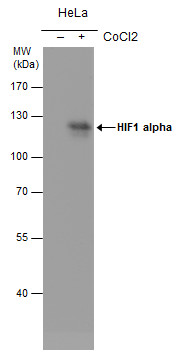
HIF1 alpha antibody [GT10211] detects HIF1 alpha protein by western blot analysis. Un-treated (-) and treated (+, 500 μM CoCl2 treatment for 24hr) HeLa whole cell extracts (30 µg) were separated by 7.5% SDS-PAGE, and the membrane was blotted with HIF1 alpha antibody [GT10211] at a dilution of 1:500. The HRP-conjugated anti-mouse IgG antibody was used to detect the primary antibody.Application
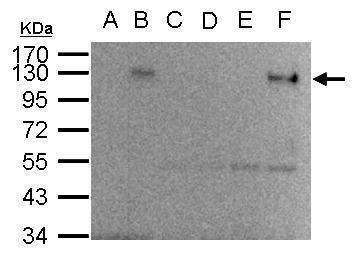
HIF-1 alpha antibody immunoprecipitates HIF-1 alpha protein in IP experiments. IP Sample: 1000 µg HepG2 whole cell lysate/extract A. 40 μg HepG2 whole cell lysate/extract from normoxia B. 40 μg HepG2 whole cell lysate/extract from hypoxia 24hr (1%O2) C. Control with 2.5 μg of preimmune mouse IgG/HepG2 whole cell lysate/extract from normoxia D. Control with 2.5 μg of preimmune mouse IgG/ HepG2 whole cell lysate/extract from hypoxia 24hr (1%O2) E. Immunoprecipitation of HIF-1 alpha protein by 2.5 μg of HIF-1 alpha antibody /HepG2 whole cell lysate/extract from normoxia F. Immunoprecipitation of HIF-1 alpha protein by 2.5 μg of HIF-1 alpha antibody /HepG2 whole cell lysate/extract from hypoxia 24hr (1%O2) 10% SDS-PAGE The immunoprecipitated HIF-1 alpha protein was detected by HIF-1 alpha antibody diluted at 1:500. EasyBlot anti-mouse IgG was used as a secondary reagent.Application
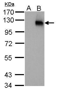
HIF1 alpha antibody [GT10211] detects HIF1A protein by western blot analysis. A. 30 µg HepG2 whole cell lysate/extract (untreated) B. 30 µg HepG2 whole cell lysate/extract (1%O2 treatment for 48hr) 7.5% SDS-PAGE HIF1 alpha antibody [GT10211] dilution: 1:250 The HRP-conjugated anti-mouse IgG antibody was used to detect the primary antibody.Application
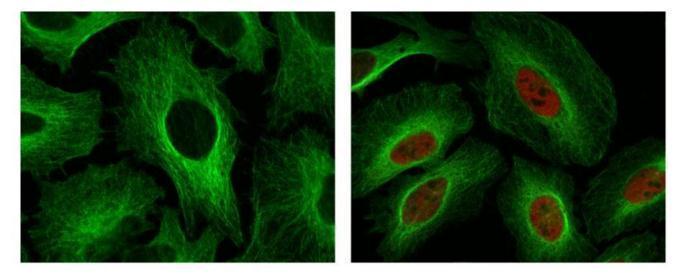
HIF1 alpha antibody detects HIF1 alpha protein at nuclear by confocal immunofluorescent analysis. Sample: Hypoxia (500μM CoCl2) treated 24hr (right) or untreated (left) HeLa cells were fixed in 4% paraformaldehyde for 15 min. Red: HIF1 alpha protein stained by HIF1 alpha antibody diluted at 1:250. Green: Alpha-tubulin, a cytoskeleton marker, stained by Rabbit Polyclonal antibody diluted at 1:500. [Images captured by Olympus FV10i Confocal Laser Scanning Microscope]| Product Name | HIF1 alpha antibody |
|---|---|
| Antibody Type | Primary Antibodies |
| Product description | Mouse Monoclonal antibody to HIF1 alpha (hypoxia-inducible factor 1-alpha)Hypoxia-inducible factor-1 (HIF1) is a transcription factor found in mammalian cells cultured under reduced oxygen tension that plays an essential role in cellular and systemic homeostatic responses to hypoxia. HIF1 is a heterodimer composed of an alpha subunit and a beta subunit. The beta subunit has been identified as the aryl hydrocarbon receptor nuclear translocator (ARNT). This gene encodes the alpha subunit of HIF-1. Overexpression of a natural antisense transcript (aHIF) of this gene has been shown to be associated with nonpapillary renal carcinomas. Two alternative transcripts encoding different isoforms have been identified. [provided by RefSeq] |
| Clonality | Monoclonal |
|---|---|
| Isotype | IgG2a |
| Clone Number | GT10211 |
| Host Species | Mouse |
| Tested Applications | ICC/IFIHC-PIPWB |
| *Optimal dilutions/concentrations should be determined by the researcher.
: |
|
| Species Reactivity | Human |
| Concentration | 1mg/ml (Please refer to the vial label for the specific concentration) |
| Purification | Affinity purified |
| Gene Synonyms | HIF-1alpha antibody HIF1 antibody HIF1-ALPHA antibody MOP1 antibody PASD8 antibody bHLHe78 antibody HIF1A antibody basic-helix-loop-helix-PAS protein MOP1 antibody class E basic helix-loop-helix protein 78 antibody ARNT-interacting protein antibod |
|---|---|
| Molecular Weight(MW) | 93 kDa (note) |
| Cellular Localization | Cytoplasm , Nucleus |
Application
HIF1 alpha antibody [GT10211] detects HIF1 alpha protein by western blot analysis. Un-treated (-) and treated (+, 500 μM CoCl2 treatment for 24hr) HeLa whole cell extracts (30 µg) were separated by 7.5% SDS-PAGE, and the membrane was blotted with HIF1 alpha antibody [GT10211] at a dilution of 1:500. The HRP-conjugated anti-mouse IgG antibody was used to detect the primary antibody.
Application
HIF-1 alpha antibody immunoprecipitates HIF-1 alpha protein in IP experiments. IP Sample: 1000 µg HepG2 whole cell lysate/extract A. 40 μg HepG2 whole cell lysate/extract from normoxia B. 40 μg HepG2 whole cell lysate/extract from hypoxia 24hr (1%O2) C. Control with 2.5 μg of preimmune mouse IgG/HepG2 whole cell lysate/extract from normoxia D. Control with 2.5 μg of preimmune mouse IgG/ HepG2 whole cell lysate/extract from hypoxia 24hr (1%O2) E. Immunoprecipitation of HIF-1 alpha protein by 2.5 μg of HIF-1 alpha antibody /HepG2 whole cell lysate/extract from normoxia F. Immunoprecipitation of HIF-1 alpha protein by 2.5 μg of HIF-1 alpha antibody /HepG2 whole cell lysate/extract from hypoxia 24hr (1%O2) 10% SDS-PAGE The immunoprecipitated HIF-1 alpha protein was detected by HIF-1 alpha antibody diluted at 1:500. EasyBlot anti-mouse IgG was used as a secondary reagent.
Application
HIF1 alpha antibody [GT10211] detects HIF1A protein by western blot analysis. A. 30 µg HepG2 whole cell lysate/extract (untreated) B. 30 µg HepG2 whole cell lysate/extract (1%O2 treatment for 48hr) 7.5% SDS-PAGE HIF1 alpha antibody [GT10211] dilution: 1:250 The HRP-conjugated anti-mouse IgG antibody was used to detect the primary antibody.
Application
HIF1 alpha antibody detects HIF1 alpha protein at nuclear by confocal immunofluorescent analysis. Sample: Hypoxia (500μM CoCl2) treated 24hr (right) or untreated (left) HeLa cells were fixed in 4% paraformaldehyde for 15 min. Red: HIF1 alpha protein stained by HIF1 alpha antibody diluted at 1:250. Green: Alpha-tubulin, a cytoskeleton marker, stained by Rabbit Polyclonal antibody diluted at 1:500. [Images captured by Olympus FV10i Confocal Laser Scanning Microscope]| Positive Control | HepG2 (hypoxia:1%O2 48hr) |
|---|---|
| Application Notes | *Optimal dilutions/concentrations should be determined by the researcher.
: |
| Form | Liquid |
|---|---|
| Storage Instructions | Keep as concentrated solution. Aliquot and store at -20ºC or below. Avoid multiple freeze-thaw cycles. |
| Storage Buffer | PBS |


