Application
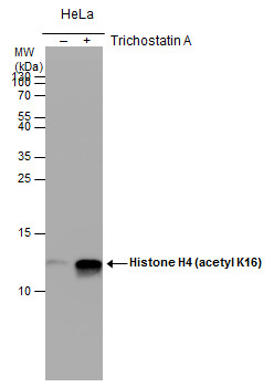
Histone H4 (acetyl Lys16) antibody detects Histone H4 (acetyl Lys16) protein by western blot analysis. Un-treated (-) and treated (+, 0.4uM Trichostatin A treatment for 18hr) HeLa whole cell extracts (30 μg) were separated by 15% SDS-PAGE, and the membrane was blotted with Histone H4 (acetyl Lys16) antibody at a dilution of 1:1000.Application
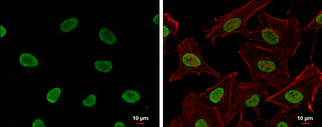
Histone H4 (acetyl Lys16) antibody [GT1271] detects Histone H4 (acetyl Lys16) protein at nucleus by immunofluorescent analysis. Sample: HeLa cells were fixed in 4% paraformaldehyde at RT for 15 min. Green: Histone H4 (acetyl Lys16) protein stained by Histone H4 (acetyl Lys16) antibody [GT1271] diluted at 1:500. Red: phalloidin, a cytoskeleton marker, diluted at 1:200. Blue: Hoechst 33342 staining. Scale bar = 10 μm.Application
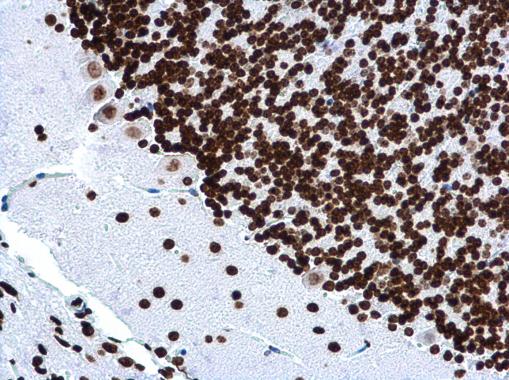
Histone H4 (acetyl Lys16) antibody [GT1271] detects Histone H4 (acetyl Lys16) protein at nucleus in mouse brain by immunohistochemical analysis. Sample: Paraffin-embedded mouse brain. Histone H4 (acetyl Lys16) antibody [GT1271] diluted at 1:500.Application
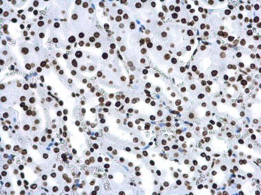
Histone H4 (acetyl Lys16) antibody [GT1271] detects Histone H4 (acetyl Lys16) protein at nucleus in mouse kidney by immunohistochemical analysis. Sample: Paraffin-embedded mouse kidney. Histone H4 (acetyl Lys16) antibody [GT1271] diluted at 1:500.| Product Name | Histone H4K16ac (Acetyl Lys16) antibody |
|---|---|
| Antibody Type | Primary Antibodies |
| Product description | Mouse Monoclonal antibody [GT1271] to Histone H4 (acetyl Lys16) (histone cluster 1, H4a)Histones are basic nuclear proteins that are responsible for the nucleosome structure of the chromosomal fiber in eukaryotes. Two molecules of each of the four core histones (H2A, H2B, H3, and H4) form an octamer, around which approximately 146 bp of DNA is wrapped in repeating units, called nucleosomes. The linker histone, H1, interacts with linker DNA between nucleosomes and functions in the compaction of chromatin into higher order structures. This gene is intronless and encodes a member of the histone H4 family. Transcripts from this gene lack polyA tails but instead contain a palindromic termination element. This gene is found in the large histone gene cluster on chromosome 6. [provided by RefSeq] |
| Immunogen | Carrier-protein conjugated synthetic peptide encompassing a sequence within the N-terminus region of human Histone H4 (acetyl Lys16). The exact sequence is proprietary. |
| Modification | Acetyl Lys16 |
| Clonality | Monoclonal |
|---|---|
| Isotype | IgG2a |
| Clone Number | GT1271 |
| Host Species | Mouse |
| Tested Applications | ICC/IFIHC-PWB |
| *Optimal dilutions/concentrations should be determined by the researcher.
: |
|
| Species Reactivity | HumanMouse |
| Predicted Reactivity | Xenopus laevisXenopus laevis (100%) |
| Concentration | 1mg/ml (Please refer to the vial label for the specific concentration) |
| Purification | Affinity purified |
| Gene Synonyms | Histone H4 acetyl Lys16 histone cluster 1 H4a HIST1H4A acetyl K16 Histone H4 acetyl Histone H4 Histone H4 acetyl K16 H4FA Histone H4 acetyl acetyl Lys16 Histone H4 |
|---|---|
| Target | Histone H4 (acetyl Lys16) |
| Molecular Weight(MW) | 11 kDa (note) |
| Cellular Localization | Nucleus |
Application
Histone H4 (acetyl Lys16) antibody detects Histone H4 (acetyl Lys16) protein by western blot analysis. Un-treated (-) and treated (+, 0.4uM Trichostatin A treatment for 18hr) HeLa whole cell extracts (30 μg) were separated by 15% SDS-PAGE, and the membrane was blotted with Histone H4 (acetyl Lys16) antibody at a dilution of 1:1000.
Application
Histone H4 (acetyl Lys16) antibody [GT1271] detects Histone H4 (acetyl Lys16) protein at nucleus by immunofluorescent analysis. Sample: HeLa cells were fixed in 4% paraformaldehyde at RT for 15 min. Green: Histone H4 (acetyl Lys16) protein stained by Histone H4 (acetyl Lys16) antibody [GT1271] diluted at 1:500. Red: phalloidin, a cytoskeleton marker, diluted at 1:200. Blue: Hoechst 33342 staining. Scale bar = 10 μm.
Application
Histone H4 (acetyl Lys16) antibody [GT1271] detects Histone H4 (acetyl Lys16) protein at nucleus in mouse brain by immunohistochemical analysis. Sample: Paraffin-embedded mouse brain. Histone H4 (acetyl Lys16) antibody [GT1271] diluted at 1:500.
Application
Histone H4 (acetyl Lys16) antibody [GT1271] detects Histone H4 (acetyl Lys16) protein at nucleus in mouse kidney by immunohistochemical analysis. Sample: Paraffin-embedded mouse kidney. Histone H4 (acetyl Lys16) antibody [GT1271] diluted at 1:500.| Positive Control | HeLa (untreated) , HeLa (0.4uM Trichostatin A for 18hr) |
|---|---|
| Application Notes | *Optimal dilutions/concentrations should be determined by the researcher.
: |
| Form | Liquid |
|---|---|
| Storage Instructions | Keep as concentrated solution. Aliquot and store at -20ºC or below. Avoid multiple freeze-thaw cycles. |
| Storage Buffer | PBS |


