Application
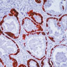
Formalin-fixed, paraffin-embedded human prostate stained with using peroxidase-conjugate and AEC chromogen. Note cytoplasmic staining of basal epithelium.| Product Name | HMW Keratin antibody |
|---|---|
| Antibody Type | Primary Antibodies |
| Product description | Mouse monoclonal [DE - SQ] to HMW Keratin |
| Immunogen | Cytoskeletal fraction prepared from human gingiva by detergent / salt extraction. |
| Clonality | Monoclonal |
|---|---|
| Isotype | IgG1 |
| Clone Number | DE-SQ |
| Host Species | Mouse |
| Tested Applications | ICC/IFIHC-P |
| Immunofluorescence: assay dependent. Immunohistochemistry (formalin fixed paraffin embedded): use at a concentration of 2 - 4μg/ml for 30 min at RT. Optimal dilutions / concentrations should be determined by the end user. Staining of formalin-fixed tissues requires digestion with Protease XXV at 1mg/ml PBS for 5 minutes at 37°C or digestion of tissue sections with pepsin at 1mg/ml Tris-HCl, pH 2.0 for 15 min at RT or 10 min at 37°C.: |
|
| Species Reactivity | HumanRat |
| Concentration | 0.2mg/ml |
| Purification | Protein G |
| Target | HMW Keratin |
|---|---|
| Tissue Specificity | This antibody recognizes cytokeratin (CK) 13, 14, 15, and 161. All squamous carcinomas and adenosquamous carcinomas are positively labeled whereas all adenocarcinomas are negative. In adenosquamous carcinomas, this antibody stains only areas with the morphologic features of squamous differentiation. Squamous carcinomas from vulva, cervix, tongue, esophagus, anus, and floor of mouth are also positive with Ab-2, either totally or focally. In contrast, adenocarcinomas of colon, cervix, parotid gland and rectum are invariably negative. In normal epithelia, DE-SQ positivity is confined to stratified epithelia, myoepithelial cells and basal cells in the prostate gland and bronchi. |
| Cellular Localization | Cytoplasmic. |
Application
Formalin-fixed, paraffin-embedded human prostate stained with using peroxidase-conjugate and AEC chromogen. Note cytoplasmic staining of basal epithelium.| Positive Control | Prostate Squamous cell carcinoma |
|---|---|
| Application Notes | Immunofluorescence: assay dependent. Immunohistochemistry (formalin fixed paraffin embedded): use at a concentration of 2 - 4μg/ml for 30 min at RT. Optimal dilutions / concentrations should be determined by the end user. Staining of formalin-fixed tissues requires digestion with Protease XXV at 1mg/ml PBS for 5 minutes at 37°C or digestion of tissue sections with pepsin at 1mg/ml Tris-HCl, pH 2.0 for 15 min at RT or 10 min at 37°C.: |
| Form | Liquid |
|---|---|
| Storage Instructions | Keep as concentrated solution, aliquot and store at 4°C. Do not freeze. |
| Storage Buffer | 10mM PBS, pH7.4, 0.2%BSA, 0.09% sodium azide |


