Application
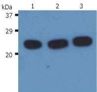
Western Blotting analysis (reducing conditions) of H-Ras in whole cell lysate using anti-H-Ras (H-RAS-03). Lane 1: HeLa human cervix carcinoma cell line. Lane 2: K567 human leukemia cell line. Lane 3: RAJI human Burkitt lymphoma cell line| Product Name | H-Ras antibody |
|---|---|
| Antibody Type | Primary Antibodies |
| Product description | Mouse Monoclonal antibody [H-RAS-03] to H-RasH-Ras is one of three ubiquitously isoforms of Ras GTPase that operate at the intracellular leaflet of the plasma membrane to regulate multiple signal transduction pathways, such as mitogen-activated protein kinase (MAPK) cascade. H Ras is anchored to the plasma membrane by farnesyl and two palmityl residues. GTP loading decreases H-Ras affinity for lipid rafts and allows the protein to target to nonraft microdomains, the primary sites of H-Ras signaling. Sos protein and other guanine nucleotide-exchange factors catalyze dissociation of GDP from Ras. Besides its roles in the plasma membrane, active H-Ras also diffuses through the cytoplasm on nanoparticles termed rasosomes, which is dependent on Ras palmitoylation. |
| Immunogen | Peptide corresponding to amino acids DIHQYREQIKRVKDSDDC of human H-Ras protein |
| Clonality | Monoclonal |
|---|---|
| Isotype | IgG1 |
| Clone Number | H-RAS-03 |
| Host Species | Mouse |
| Tested Applications | WB |
| WB: Use at 0.5 - 1 µg/mL. Positive Control:RAJI human Burkitt lymphoma cell lineHeLa human cervix carcinoma cell lineK567 human leukemia cell line Sample preparation: Resuspend approx. 50 mil. cells in 1 ml cold Lysis buffer (1% laurylmaltoside in 20 mM Tris/Cl, 100 mM NaCl pH 8.2, 50 mM NaF including Protease inhibitor Cocktail). Incubate 60 min on ice. Centrifuge to remove cell debris. Mix lysate with reducing Laemmli SDS-PAGE Sample buffer. Boil for 5 min. Use under reducing conditions: |
|
| Species Reactivity | Human |
| Concentration | 1mg/ml |
| Gene Synonyms | H Ras HRas |
|---|---|
| Target | H-Ras |
| Tissue Specificity | The antibody H-RAS-03 reacts with human H-Ras, an ubiquitously expressed 21 kDa intracellular protein. Although reactivity with other species has not been determined, it is probable as the epitope is highly conserved among animals. |
| Cellular Localization | Shuttles between the plasma membrane and the Golgi apparatus. |
Application
Western Blotting analysis (reducing conditions) of H-Ras in whole cell lysate using anti-H-Ras (H-RAS-03). Lane 1: HeLa human cervix carcinoma cell line. Lane 2: K567 human leukemia cell line. Lane 3: RAJI human Burkitt lymphoma cell line| Application Notes | WB: Use at 0.5 - 1 µg/mL. Positive Control:RAJI human Burkitt lymphoma cell lineHeLa human cervix carcinoma cell lineK567 human leukemia cell line Sample preparation: Resuspend approx. 50 mil. cells in 1 ml cold Lysis buffer (1% laurylmaltoside in 20 mM Tris/Cl, 100 mM NaCl pH 8.2, 50 mM NaF including Protease inhibitor Cocktail). Incubate 60 min on ice. Centrifuge to remove cell debris. Mix lysate with reducing Laemmli SDS-PAGE Sample buffer. Boil for 5 min. Use under reducing conditions: |
|---|
| Form | Liquid |
|---|---|
| Storage Instructions | Store at 2-8°C. Do not use after expiration date stamped on vial label. For long-term storage aliquot and store at -20°C. Avoid freeze/thaw cycles. |
| Storage Buffer | Phosphate buffered saline (PBS) with 15 mM sodium azide, approx. pH 7.4 |


