Application
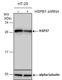
Non-transfected (–) and transfected (+) HT-29 whole cell extracts (30 µg) were separated by 12% SDS-PAGE, and the membrane was blotted with HSP27 antibody diluted at 1:10000. The HRP-conjugated anti-rabbit IgG antibody was used to detect the primary antibody.Application
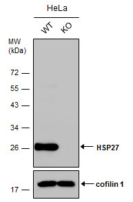
Wild-type (WT) and HSP27 knownout (KO) HeLa cell extracts (30 µg) were separated by 12% SDS-PAGE, and the membrane was blotted with HSP27 antibody diluted at 1:20000. The HRP-conjugated anti-rabbit IgG antibody was used to detect the primary antibody.Application
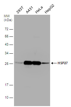
Various whole cell extracts (30 µg) were separated by 12% SDS-PAGE, and the membrane was blotted with HSP27 antibody diluted at 1:10000. The HRP-conjugated anti-rabbit IgG antibody was used to detect the primary antibody.Application
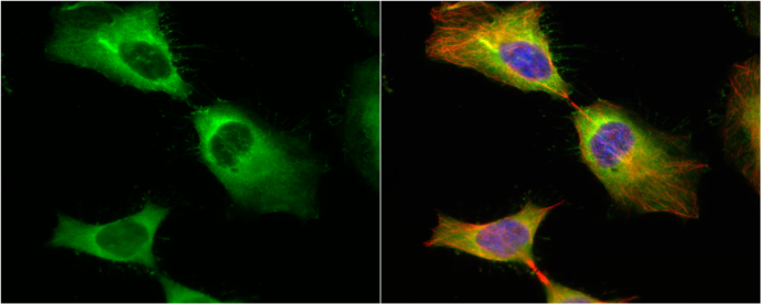
HSP27 antibody detects HSP27 protein at cytoplasm and cytoskeleton by immunofluorescent analysis. Sample: HeLa cells were fixed in 4% paraformaldehyde at RT for 15 min. Green: HSP27 protein stained by HSP27 antibody diluted at 1:1000. Red: phalloidin, a cytoskeleton marker, stained by phalloidin (invitrogen, A12380) diluted at 1:200. Blue: Hoechst 33342 staining.Application
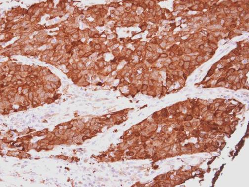
HSP27 antibody detects HSP27 protein at cytoplasm in human lung papillary carcinoma by immunohistochemical analysis. Sample: Paraffin-embedded human lung papillary carcinoma. HSP27 antibody diluted at 1:500.Application
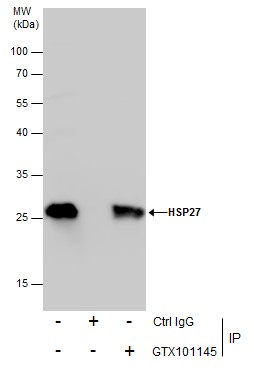
Immunoprecipitation of HSP27 protein from HeLa whole cell extracts using 5 μg of HSP27 antibody . Western blot analysis was performed using HSP27 antibody . EasyBlot anti-Rabbit IgG was used as a secondary reagent.Application
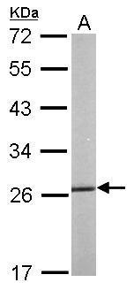
Sample (50 µg of whole cell lysate) A: mouse liver 12% SDS PAGE diluted at 1:1000 The HRP-conjugated anti-rabbit IgG antibody was used to detect the primary antibody.| Product Name | HSP27 antibody |
|---|---|
| Antibody Type | Primary Antibodies |
| Product description | Rabbit Polyclonal antibody to HSP27 (heat shock 27kDa protein 1)The protein encoded by this gene is induced by environmental stress and developmental changes. The encoded protein is involved in stress resistance and actin organization and translocates from the cytoplasm to the nucleus upon stress induction. Defects in this gene are a cause of Charcot-Marie-Tooth disease type 2F (CMT2F) and distal hereditary motor neuropathy (dHMN). [provided by RefSeq] |
| Immunogen | Recombinant protein encompassing a sequence within the center region of human HSP27. The exact sequence is proprietary. |
| Clonality | Polyclonal |
|---|---|
| Isotype | IgG |
| Host Species | Rabbit |
| Tested Applications | FACSICC/IFIHC-PIPWB |
| *Optimal dilutions/concentrations should be determined by the researcher.
: |
|
| Species Reactivity | HumanMouseRat |
| Predicted Reactivity | Dog, Pig, Rhesus Monkey, BovineDog (86%), Pig (86%), Rhesus Monkey (100%), Bovine (83%) |
| Concentration | 1mg/ml |
| Purification | Affinity purified |
| Gene Synonyms | CMT2F antibody DKFZp586P1322 antibody HMN2B antibody HS.76067 antibody HSP27 antibody HSP28 antibody Hsp25 antibody SRP27 antibody HSPB1 antibody 28 kDa heat shock protein antibody stress-responsive protein 27 antibody heat shock protein beta-1 |
|---|---|
| Molecular Weight(MW) | 23 kDa (note) |
| Cellular Localization | Cytoplasm , Nucleus , Spindle |
Application
Non-transfected (–) and transfected (+) HT-29 whole cell extracts (30 µg) were separated by 12% SDS-PAGE, and the membrane was blotted with HSP27 antibody diluted at 1:10000. The HRP-conjugated anti-rabbit IgG antibody was used to detect the primary antibody.
Application
Wild-type (WT) and HSP27 knownout (KO) HeLa cell extracts (30 µg) were separated by 12% SDS-PAGE, and the membrane was blotted with HSP27 antibody diluted at 1:20000. The HRP-conjugated anti-rabbit IgG antibody was used to detect the primary antibody.
Application
Various whole cell extracts (30 µg) were separated by 12% SDS-PAGE, and the membrane was blotted with HSP27 antibody diluted at 1:10000. The HRP-conjugated anti-rabbit IgG antibody was used to detect the primary antibody.
Application
HSP27 antibody detects HSP27 protein at cytoplasm and cytoskeleton by immunofluorescent analysis. Sample: HeLa cells were fixed in 4% paraformaldehyde at RT for 15 min. Green: HSP27 protein stained by HSP27 antibody diluted at 1:1000. Red: phalloidin, a cytoskeleton marker, stained by phalloidin (invitrogen, A12380) diluted at 1:200. Blue: Hoechst 33342 staining.
Application
HSP27 antibody detects HSP27 protein at cytoplasm in human lung papillary carcinoma by immunohistochemical analysis. Sample: Paraffin-embedded human lung papillary carcinoma. HSP27 antibody diluted at 1:500.
Application
Immunoprecipitation of HSP27 protein from HeLa whole cell extracts using 5 μg of HSP27 antibody . Western blot analysis was performed using HSP27 antibody . EasyBlot anti-Rabbit IgG was used as a secondary reagent.
Application
Sample (50 µg of whole cell lysate) A: mouse liver 12% SDS PAGE diluted at 1:1000 The HRP-conjugated anti-rabbit IgG antibody was used to detect the primary antibody.| Positive Control | 293T , A431 , HeLa , HepG2 , HT-29 , Mouse liver |
|---|---|
| Application Notes | *Optimal dilutions/concentrations should be determined by the researcher.
: |
| Form | Liquid |
|---|---|
| Storage Instructions | Keep as concentrated solution. Aliquot and store at -20ºC or below. Avoid multiple freeze-thaw cycles. |
| Storage Buffer | 1XPBS, 20% Glycerol (pH7). 0.025% ProClin 300 was added as a preservative. |


