Application
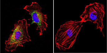
Immunofluorescent analysis of Heat Shock Protein 60 using Heat Shock Protein 60 Monoclonal Antibody shows staining in BEAS-2B Cells. Heat Shock Protein 60 (green), F-Actin staining with Phalloidin (red) and nuclei with DAPI (blue) is shown. Cells were grown on chamber slides and fixed with formaldehyde prior to staining. Cells were probed without (control) or with an antibody recognizing Heat Shock Protein 60 at a dilution of 1:100 over night at 4 C, washed with PBS and incubated with a DyLight-488 conjugated secondary antibody.Application
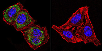
Immunofluorescent analysis of Heat Shock Protein 60 using Heat Shock Protein 60 Monoclonal Antibody shows staining in Hela Cells. Heat Shock Protein 60 (green), F-Actin staining with Phalloidin (red) and nuclei with DAPI (blue) is shown. Cells were grown on chamber slides and fixed with formaldehyde prior to staining. Cells were probed without (control) or with an antibody recognizing Heat Shock Protein 60 at a dilution of 1:100 over night at 4 C, washed with PBS and incubated with a DyLight-488 conjugated secondary antibody.Application
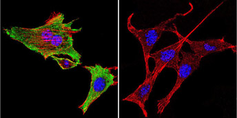
Immunofluorescent analysis of Heat Shock Protein 60 using Heat Shock Protein 60 Monoclonal Antibody shows staining in 293Cells. Heat Shock Protein 60 (green), F-Actin staining with Phalloidin (red) and nuclei with DAPI (blue) is shown. Cells were grown on chamber slides and fixed with formaldehyde prior to staining. Cells were probed without (control) or with an antibody recognizing Heat Shock Protein 60 at a dilution of 1:20 over night at 4 C, washed with PBS and incubated with a DyLight-488 conjugated secondary antibody.| Product Name | HSP60 antibody |
|---|---|
| Antibody Type | Primary Antibodies |
| Product description | Mouse Monoclonal antibody to HSP60Heat shock proteins (HSP) are expressed in response to various biological stresses, including high temperatures. There are several major families of HSPs including HSP70, HSP90 and HSP100. HSP60, an ~60 kDa protein, is the human homologue of HSP65 (a major antigenic mycobacterial protein) and E. coli GroEL. HSP60, also referred to as chaperonin-60, P1 & mitonin, has been classified as a member of a family of proteins termed chaperonins which act to recognize and stabilize polypeptide intermediates during folding, assembly and disassembly. HSP60 is a constitutively expressed mitochondrial protein that shares many characteristics with HSP70 including high abundance, induction by environmental stress, ATPase activity and a critical role in protein assembly and folding. However, in contrast with HSP70, HSP60 functions as a 14 subunit oligomer which is thought to interact with only 1 or 2 target polypeptides at one time. |
| Immunogen | Recombinant Chlamydia trachomatis HSP60 protein. |
| Clonality | Monoclonal |
|---|---|
| Isotype | IgG1 |
| Clone Number | A57-B9 |
| Host Species | Mouse |
| Tested Applications | ELISAICC/IFIHCIPWB |
| ICC/IF: 1:250: |
|
| Species Reactivity | Bacteria |
| Concentration | 1mg/ml |
| Purification | Affinity purified |
| Gene Synonyms | groEL |
|---|---|
| Target | Heat Shock Protein 60 |
Application
Immunofluorescent analysis of Heat Shock Protein 60 using Heat Shock Protein 60 Monoclonal Antibody shows staining in BEAS-2B Cells. Heat Shock Protein 60 (green), F-Actin staining with Phalloidin (red) and nuclei with DAPI (blue) is shown. Cells were grown on chamber slides and fixed with formaldehyde prior to staining. Cells were probed without (control) or with an antibody recognizing Heat Shock Protein 60 at a dilution of 1:100 over night at 4 C, washed with PBS and incubated with a DyLight-488 conjugated secondary antibody.
Application
Immunofluorescent analysis of Heat Shock Protein 60 using Heat Shock Protein 60 Monoclonal Antibody shows staining in Hela Cells. Heat Shock Protein 60 (green), F-Actin staining with Phalloidin (red) and nuclei with DAPI (blue) is shown. Cells were grown on chamber slides and fixed with formaldehyde prior to staining. Cells were probed without (control) or with an antibody recognizing Heat Shock Protein 60 at a dilution of 1:100 over night at 4 C, washed with PBS and incubated with a DyLight-488 conjugated secondary antibody.
Application
Immunofluorescent analysis of Heat Shock Protein 60 using Heat Shock Protein 60 Monoclonal Antibody shows staining in 293Cells. Heat Shock Protein 60 (green), F-Actin staining with Phalloidin (red) and nuclei with DAPI (blue) is shown. Cells were grown on chamber slides and fixed with formaldehyde prior to staining. Cells were probed without (control) or with an antibody recognizing Heat Shock Protein 60 at a dilution of 1:20 over night at 4 C, washed with PBS and incubated with a DyLight-488 conjugated secondary antibody.| Application Notes | ICC/IF: 1:250: |
|---|
| Form | Liquid |
|---|---|
| Storage Instructions | Keep as concentrated solution. Aliquot and store at -20ºC or below. Avoid multiple freeze-thaw cycles. |
| Storage Buffer | PBS containing 0.05% sodium azide. |


