Application
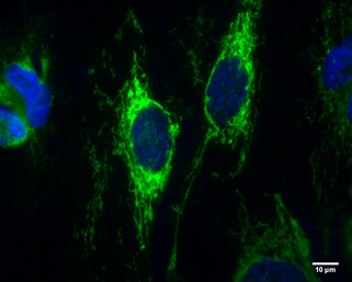
IDH2 antibody [GT673] detects IDH2 protein at Mitochondria by immunofluorescent analysis. Sample: HeLa cells were fixed in 4% paraformaldehyde at RT for 10 min. Green: IDH2 protein stained by IDH2 antibody [GT673] diluted at 1:100. Blue: Hoechst 33342 staining. Scale bar = 10 μm.Application
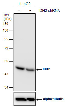
Non-transfected (–) and transfected (+) HepG2 whole cell extracts (30 µg) were separated by 10% SDS-PAGE, and the membrane was blotted with IDH2 antibody [GT673] diluted at 1:500. The HRP-conjugated anti-mouse IgG antibody was used to detect the primary antibody.Application
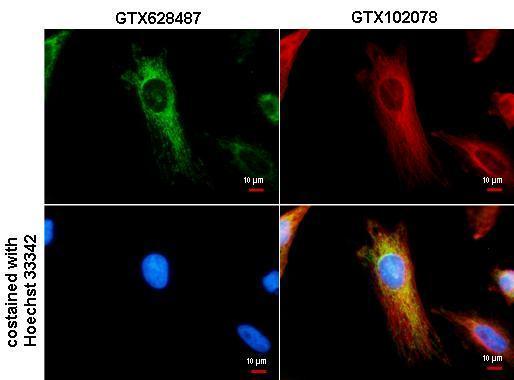
IDH2 antibody detects IDH2 protein at Mitochondria by immunofluorescent analysis. Sample: HeLa cells were fixed in 4% paraformaldehyde/PBS for 15 min. Green: IDH2 protein stained by IDH2 antibody diluted at 1:500. Red: alpha Tubulin protein stained by alpha Tubulin antibody diluted at 1:500. Blue: Hoechst 33342 staining.Application
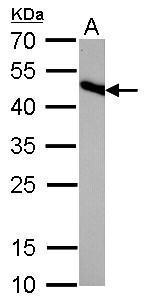
IDH2 antibody [GT673] detects IDH2 protein by western blot analysis. A. 50 µg rat brain lysate/extract 12% SDS-PAGE IDH2 antibody [GT673] dilution: 1:1000 The HRP-conjugated anti-mouse IgG antibody was used to detect the primary antibody.Application
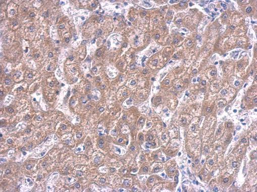
IDH2 antibody detects IDH2 protein at cytosol on human hepatoma by immunohistochemical analysis. Sample: Paraffin-embedded hepatoma. IDH2 antibody dilution: 1:100.Application
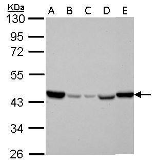
IDH2 antibody detects IDH2 protein by western blot analysis. A. 30 µg Jurkat whole cell lysate/extract B. 30 µg Raji whole cell lysate/extract C. 30 µg K562 whole cell lysate/extract D. 30 µg THP-1 whole cell lysate/extract E. 30 µg NCI-H929 whole cell lysate/extract 10% SDS-PAGE IDH2 antibody dilution: 1:1000 The HRP-conjugated anti-mouse IgG antibody was used to detect the primary antibody.Application
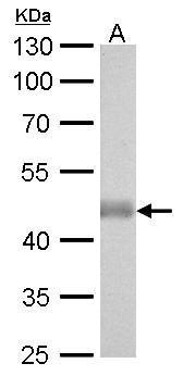
IDH2 antibody [GT673] detects IDH2 protein by western blot analysis. A. 50 µg mouse brain lysate/extract 10% SDS-PAGE IDH2 antibody [GT673] dilution: 1:1000 The HRP-conjugated anti-mouse IgG antibody was used to detect the primary antibody.| Product Name | IDH2 antibody [GT673] |
|---|---|
| Antibody Type | Primary Antibodies |
| Product description | Mouse Monoclonal antibody to IDH2 (isocitrate dehydrogenase 2 (NADP+), mitochondrial)Isocitrate dehydrogenases catalyze the oxidative decarboxylation of isocitrate to 2-oxoglutarate. These enzymes belong to two distinct subclasses, one of which utilizes NAD(+) as the electron acceptor and the other NADP(+). Five isocitrate dehydrogenases have been reported: three NAD(+)-dependent isocitrate dehydrogenases, which localize to the mitochondrial matrix, and two NADP(+)-dependent isocitrate dehydrogenases, one of which is mitochondrial and the other predominantly cytosolic. Each NADP(+)-dependent isozyme is a homodimer. The protein encoded by this gene is the NADP(+)-dependent isocitrate dehydrogenase found in the mitochondria. It plays a role in intermediary metabolism and energy production. This protein may tightly associate or interact with the pyruvate dehydrogenase complex. [provided by RefSeq] |
| Immunogen | Recombinant protein encompassing a sequence within the center region of human IDH2. The exact sequence is proprietary. |
| Clonality | Monoclonal |
|---|---|
| Isotype | IgG1 |
| Clone Number | GT673 |
| Host Species | Mouse |
| Tested Applications | ICC/IF,IHC-P,WB |
| *Optimal dilutions/concentrations should be determined by the researcher. | |
| Species Reactivity | Human,Mouse,Rat |
| Predicted Reactivity | Zebrafish, Xenopus laevis, Pig, Chicken, Rhesus Monkey, BovineZebrafish (89%), Xenopus laevis (91%), Pig (96%), Chicken (95%), Rhesus Monkey (100%), Bovine (96%) |
| Concentration | 1mg/ml (Please refer to the vial label for the specific concentration) |
| Purification | Affinity purified |
| Gene Synonyms | D2HGA2 antibody, ICD-M antibody, IDH antibody, IDHM antibody, IDP antibody, IDPM antibody, mNADP-IDH antibody, IDH2 antibody, NADP(+)-specific ICDH antibody, isocitrate dehydrogenase [NADP], mitochondrial antibody, oxalosuccinate decarboxylase antibody, is |
|---|---|
| Molecular Weight(MW) | 51 kDa (note) (2) |
| Cellular Localization | Mitochondrion |
Application
IDH2 antibody [GT673] detects IDH2 protein at Mitochondria by immunofluorescent analysis. Sample: HeLa cells were fixed in 4% paraformaldehyde at RT for 10 min. Green: IDH2 protein stained by IDH2 antibody [GT673] diluted at 1:100. Blue: Hoechst 33342 staining. Scale bar = 10 μm.
Application
Non-transfected (–) and transfected (+) HepG2 whole cell extracts (30 µg) were separated by 10% SDS-PAGE, and the membrane was blotted with IDH2 antibody [GT673] diluted at 1:500. The HRP-conjugated anti-mouse IgG antibody was used to detect the primary antibody.
Application
IDH2 antibody detects IDH2 protein at Mitochondria by immunofluorescent analysis. Sample: HeLa cells were fixed in 4% paraformaldehyde/PBS for 15 min. Green: IDH2 protein stained by IDH2 antibody diluted at 1:500. Red: alpha Tubulin protein stained by alpha Tubulin antibody diluted at 1:500. Blue: Hoechst 33342 staining.
Application
IDH2 antibody [GT673] detects IDH2 protein by western blot analysis. A. 50 µg rat brain lysate/extract 12% SDS-PAGE IDH2 antibody [GT673] dilution: 1:1000 The HRP-conjugated anti-mouse IgG antibody was used to detect the primary antibody.
Application
IDH2 antibody detects IDH2 protein at cytosol on human hepatoma by immunohistochemical analysis. Sample: Paraffin-embedded hepatoma. IDH2 antibody dilution: 1:100.
Application
IDH2 antibody detects IDH2 protein by western blot analysis. A. 30 µg Jurkat whole cell lysate/extract B. 30 µg Raji whole cell lysate/extract C. 30 µg K562 whole cell lysate/extract D. 30 µg THP-1 whole cell lysate/extract E. 30 µg NCI-H929 whole cell lysate/extract 10% SDS-PAGE IDH2 antibody dilution: 1:1000 The HRP-conjugated anti-mouse IgG antibody was used to detect the primary antibody.
Application
IDH2 antibody [GT673] detects IDH2 protein by western blot analysis. A. 50 µg mouse brain lysate/extract 10% SDS-PAGE IDH2 antibody [GT673] dilution: 1:1000 The HRP-conjugated anti-mouse IgG antibody was used to detect the primary antibody.| Application Notes | *Optimal dilutions/concentrations should be determined by the researcher. |
|---|
| Form | Liquid |
|---|---|
| Storage Instructions | Keep as concentrated solution. Aliquot and store at -20ºC or below. Avoid multiple freeze-thaw cycles. |
| Storage Buffer | PBS |


