Application
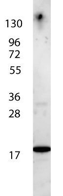
GeneTex's anti-Human IL-7 antibody shows detection of a band ~17 kDa in size corresponding to recombinant human IL-7. The identity of the faint higher molecular weight band may represent a homodimer. Molecular weight markers are also shown (left). After transfer, the membrane was blocked overnight with 3% BSA in TBS followed by reaction with primary antibody at a 1:1,000 dilution. Detection occurred using peroxidase conjugated anti-Rabbit IgG secondary antibody diluted 1:40,000 in blocking buffer) for 30 min at RT followed by reaction with FemtoMax chemiluminescent substrate. Image was captured using VersaDoc MP 4000 imaging system (Bio-Rad).| Product Name | IL7 antibody |
|---|---|
| Antibody Type | Primary Antibodies |
| Product description | Rabbit Polyclonal antibody to IL-7Interleukin 7 (IL7) is a lymphoid cell growth factor that affects pre-B, pro-B, and early T cells. IL7 was previously known as pre-B cell growth factor and lymphopoietin 1. IL7 supports the growth of early B cells from long-term lymphoid bone marrow cultures. It is mitogenic to thymocytes and enhances the response of cells to other stimuli such as polyhydroxyalkanoate (PHA) and concanavalin A (ConA). IL7 stimulates the proliferation of CD4+/CD8+ cells. The proliferative response of thymocytes to IL7 is not affected by antibodies to the T cell growth factors such as IL2, IL4 and IL6, suggesting that IL7 is capable of stimulating T cell proliferation through a pathway independent of the known T cell growth factors. Mature T cells respond to IL7 and Con A, but not to IL7 alone. In myeloid lineage cells, IL7 upregulates the production of pro-inflammatory cytokines and stimulates the tumoricidal activity of monocytes/macrophages. IL7 is expressed by adherent stromal cells from various tissues. |
| Immunogen | This purified antibody was prepared from whole rabbit serum produced by repeated immunizations with full length recombinant human IL-7 protein. |
| Clonality | Polyclonal |
|---|---|
| Isotype | IgG |
| Host Species | Rabbit |
| Tested Applications | ELISAWB |
| *Optimal dilutions/concentrations should be determined by the researcher.
: |
|
| Species Reactivity | Human |
| Concentration | 1mg/ml (Please refer to the vial label for the specific concentration) |
| Gene Synonyms | IL7 |
|---|---|
| Target | IL-7 |
Application
GeneTex's anti-Human IL-7 antibody shows detection of a band ~17 kDa in size corresponding to recombinant human IL-7. The identity of the faint higher molecular weight band may represent a homodimer. Molecular weight markers are also shown (left). After transfer, the membrane was blocked overnight with 3% BSA in TBS followed by reaction with primary antibody at a 1:1,000 dilution. Detection occurred using peroxidase conjugated anti-Rabbit IgG secondary antibody diluted 1:40,000 in blocking buffer) for 30 min at RT followed by reaction with FemtoMax chemiluminescent substrate. Image was captured using VersaDoc MP 4000 imaging system (Bio-Rad).| Application Notes | *Optimal dilutions/concentrations should be determined by the researcher.
: |
|---|
| Form | Liquid |
|---|---|
| Storage Instructions | Store vial at 4° C for short-term use. For extended storage aliquot contents and freeze at -20° C or below. Avoid cycles of freezing and thawing. |
| Storage Buffer | 0.02 M Potassium Phosphate, 0.15 M Sodium Chloride, pH 7.2 |


