Application
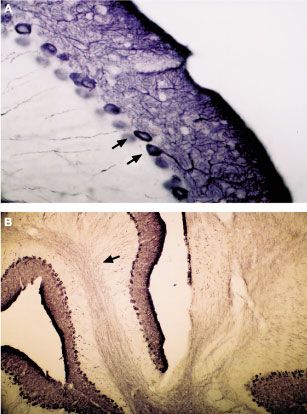
IHC analysis of mouse cerebellum using IP3 Receptor 1 antibody. Immunoreactivity appears in Purkinje cells (arrows in A) and their dendritic trees. In addition, axonal processes coarsing through cerebellar white matter are visualized (arrow in B).Application
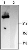
WB analysis of rat brain lysates using IP3 Receptor 1 antibody at a dilution of 1:2000 in the (1) absence or (2) presence of blocking peptide.| Product Name | IP3 Receptor I antibody |
|---|---|
| Antibody Type | Primary Antibodies |
| Product description | Rabbit Polyclonal antibody to IP3 Receptor 1This gene belongs to the Insp3 receptor family. It encodes an intracellular receptor for inositol 1,4,5-trisphosphate. Upon stimulation by inositol 1,4,5-trisphosphate, this receptor mediates calcium release from the endoplasmic reticulum. Alternatively spliced transcript variants encoding distinct isoforms have been found for this gene. [provided by RefSeq, Jul 2012] |
| Immunogen | Peptide (C)RIGLLGHPPHMNVNPQQPA, corresponding to amino acid residues 2732-2750 of rat IP3 Receptor-1 (Accession P29994). Intracellular, C-terminus. |
| Clonality | Polyclonal |
|---|---|
| Host Species | Rabbit |
| Tested Applications | ICC/IFIHCIPWB |
| ICC/IF:1:200~1:500 IHC:1:50~1:200 IP:1:100~1:500 WB:1:500~1:2000 Notes:Optimal dilutions/concentrations should be determined by the researcher. |
|
| Species Reactivity | HumanMouseRat |
| Concentration | 1mg/ml |
| Purification | Protein A |
| Gene Synonyms | InsP3R Itpr1 InsP3R1 I145TR inositol 1 4 5-trisphosphate receptor type 1 |
|---|---|
| Target | IP3 Receptor 1 |
Application
IHC analysis of mouse cerebellum using IP3 Receptor 1 antibody. Immunoreactivity appears in Purkinje cells (arrows in A) and their dendritic trees. In addition, axonal processes coarsing through cerebellar white matter are visualized (arrow in B).
Application
WB analysis of rat brain lysates using IP3 Receptor 1 antibody at a dilution of 1:2000 in the (1) absence or (2) presence of blocking peptide.| Application Notes | ICC/IF:1:200~1:500 IHC:1:50~1:200 IP:1:100~1:500 WB:1:500~1:2000 Notes:Optimal dilutions/concentrations should be determined by the researcher. |
|---|
| Form | Liquid |
|---|---|
| Storage Instructions | Keep as concentrated solution. Aliquot and store at -20ºC or below. Avoid multiple freeze-thaw cycles. |
| Storage Buffer | Phosphate buffered saline (PBS), pH 7.4, 1% BSA and 0.05% NaN3. |


