Application
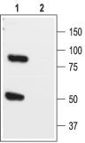
Anti-K2P1.1_(TWIK-1)_(extracellular) - Western blot analysis of rat brain lysate: 1. Anti-K2P1.1 (TWIK-1) (extracellular) antibody, (1:200). 2. Anti-K2P1.1 (TWIK-1) (extracellular) antibody, preincubated with the control peptide antigen.Application
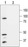
Anti-K2P1.1_(TWIK-1)_(extracellular) - Western blot analysis of HEK-293-K2P1.1 transfected cells: 1. Anti-K2P1.1 (TWIK-1) (extracellular) antibody, (1:300). 2. Anti-K2P1.1 (TWIK-1) (extracellular) antibody, preincubated with the control peptide antigen.Application
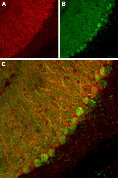
Anti-K2P1.1_(TWIK-1)_(extracellular) - Expression of K2P1.1 in mouse cerebellum Immunohistochemical staining of mouse cerebellum using Anti-K2P1.1 (TWIK-1) (extracellular) antibody. A. K2P1.1 channel appears in glial processes (red) . B. staining of Purkinje nerve cells with mouse anti calbindin D28K (a calcium binding protein, green). C. Confocal merge of K2P1.1 channel and calbindin D28K demonstrates the separate localization of these proteins.| Product Name | KCNK1 antibody |
|---|---|
| Antibody Type | Primary Antibodies |
| Product description | Rabbit Polyclonal antibody to KCNK1This gene encodes one of the members of the superfamily of potassium channel proteins containing two pore-forming P domains. The product of this gene has not been shown to be a functional channel, however, it may require other non-pore-forming proteins for activity. [provided by RefSeq, Jul 2008] |
| Immunogen | Peptide RQELRKLKRRFLEEHEC, corresponding to amino acid residues 53-69 of human K2P1.1 (Accession O00180). Extracellular, near the P1 loop. |
| Clonality | Polyclonal |
|---|---|
| Isotype | IgG |
| Host Species | Rabbit |
| Tested Applications | IHCWB |
| IHC:1:50~1:200 WB:1:500~1:2000 Notes:Optimal dilutions/concentrations should be determined by the researcher. |
|
| Species Reactivity | HumanMouseRat |
| Concentration | 1mg/ml |
| Purification | Affinity purified |
| Gene Synonyms | potassium channel subfamily K member 1 HOHO DPK KCNO1 TWIK-1 K2p1.1 TWIK1 K2P1 |
|---|---|
| Target | KCNK1 |
Application
Anti-K2P1.1_(TWIK-1)_(extracellular) - Western blot analysis of rat brain lysate: 1. Anti-K2P1.1 (TWIK-1) (extracellular) antibody, (1:200). 2. Anti-K2P1.1 (TWIK-1) (extracellular) antibody, preincubated with the control peptide antigen.
Application
Anti-K2P1.1_(TWIK-1)_(extracellular) - Western blot analysis of HEK-293-K2P1.1 transfected cells: 1. Anti-K2P1.1 (TWIK-1) (extracellular) antibody, (1:300). 2. Anti-K2P1.1 (TWIK-1) (extracellular) antibody, preincubated with the control peptide antigen.
Application
Anti-K2P1.1_(TWIK-1)_(extracellular) - Expression of K2P1.1 in mouse cerebellum Immunohistochemical staining of mouse cerebellum using Anti-K2P1.1 (TWIK-1) (extracellular) antibody. A. K2P1.1 channel appears in glial processes (red) . B. staining of Purkinje nerve cells with mouse anti calbindin D28K (a calcium binding protein, green). C. Confocal merge of K2P1.1 channel and calbindin D28K demonstrates the separate localization of these proteins.| Application Notes | IHC:1:50~1:200 WB:1:500~1:2000 Notes:Optimal dilutions/concentrations should be determined by the researcher. |
|---|
| Form | Liquid |
|---|---|
| Storage Instructions | Keep as concentrated solution. Aliquot and store at -20ºC or below. Avoid multiple freeze-thaw cycles. |
| Storage Buffer | Phosphate buffered saline (PBS), pH 7.4, 1% BSA, 0.025% NaN3. |


