Application
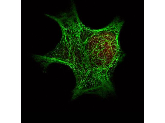
Immunofluorescence Microscopy of GeneTex Anti-Keratin antibody was used with GeneTex Dylight 488 goat anti-mouse (shown in green) to detect Keratin by Immunofluorescence. In the same experiment, GeneTex polyclonal Anti-HDAC-1 antibody was used with Anti-Rabbit IgG (shown in red) to detect HDAC-1.Application
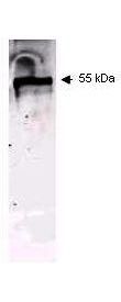
Western blot using GeneTex Immunochemical's Mouse Mab-anti-Keratin antibody. This antibody recognizes a single 56 kDa band corresponding to human keratin as confirmed by the position of molecular weight markers (not shown). Approximatley 100 µg of keratin from human epidermis was applied under reducing conditions. 1:400 dilution of Mab anti-Keratin was used for 2h followed by detection using a 1:5,000 dilution of IRDyeTM800 conjugated Goat anti-Mouse IgG [H&L] and visualization using the Odyssey® Infrared Imaging System developed by LI-COR.Application
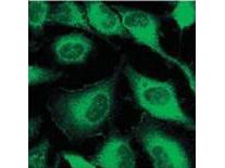
Immunofluorescence microscopy using GeneTex Immunochemical's Mouse Mab-anti-Keratin antibody. Confocal slices of HeLa cells are between 0.5 and 0.6 um where the image is taken near the bottom of the cell. Use FITC a 1:2,000 dilution of FITC-conjugated Goat anti-Mouse IgG [H&L] for detection.| Product Name | Keratin antibody [C11] |
|---|---|
| Antibody Type | Primary Antibodies |
| Product description | Mouse Monoclonal antibody [C11] to KeratinCytokeratins (CK) are intermediate filaments of epithelial cells, both in keratinizing tissue (i.e. skin) and non-keratinizing cells (i.e. mesothelial) |
| Immunogen | This protein A purified monoclonal antibody was produced by repeated immunizations with purified human cytoskeletal preparations from A431 cells. |
| Clonality | Monoclonal |
|---|---|
| Isotype | IgG1 |
| Clone Number | C11 |
| Host Species | Mouse |
| Tested Applications | ELISA,ICC/IF,IHC,IHC-P,IP,WB |
| *Optimal dilutions/concentrations should be determined by the researcher. | |
| Species Reactivity | Human |
| Concentration | 1mg/ml |
| Purification | Protein A |
| Gene Synonyms | KRT6E , KRT6C , K6E |
|---|---|
| Target | Keratin |
| Tissue Specificity | This antibody reacts with keratin (56 kDa), keratin 17 (46 kDa), keratin 18 (45 kDa) and keratin 19 (40 kDa) derived from humans. |
Application
Immunofluorescence Microscopy of GeneTex Anti-Keratin antibody was used with GeneTex Dylight 488 goat anti-mouse (shown in green) to detect Keratin by Immunofluorescence. In the same experiment, GeneTex polyclonal Anti-HDAC-1 antibody was used with Anti-Rabbit IgG (shown in red) to detect HDAC-1.
Application
Western blot using GeneTex Immunochemical's Mouse Mab-anti-Keratin antibody. This antibody recognizes a single 56 kDa band corresponding to human keratin as confirmed by the position of molecular weight markers (not shown). Approximatley 100 µg of keratin from human epidermis was applied under reducing conditions. 1:400 dilution of Mab anti-Keratin was used for 2h followed by detection using a 1:5,000 dilution of IRDyeTM800 conjugated Goat anti-Mouse IgG [H&L] and visualization using the Odyssey® Infrared Imaging System developed by LI-COR.
Application
Immunofluorescence microscopy using GeneTex Immunochemical's Mouse Mab-anti-Keratin antibody. Confocal slices of HeLa cells are between 0.5 and 0.6 um where the image is taken near the bottom of the cell. Use FITC a 1:2,000 dilution of FITC-conjugated Goat anti-Mouse IgG [H&L] for detection.| Application Notes | *Optimal dilutions/concentrations should be determined by the researcher. |
|---|
| Form | Liquid |
|---|---|
| Storage Instructions | Store vial at -20° C prior to opening. Aliquot contents and freeze at -20° C or below for extended storage. Avoid cycles of freezing and thawing. |
| Storage Buffer | 0.02 M Potassium Phosphate, 0.15 M Sodium Chloride, pH 7.2 and 0.01% (w/v) Sodium Azide |


