Application
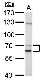
Lamin A + C antibody detects Lamin A + C protein by Western blot analysis. A. 30 μg NIH-3T3 whole cell lysate/extract 7.5 % SDS-PAGE Lamin A + C antibody dilution: 1:1000Application
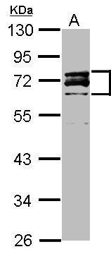
Sample (20 ug ) A: HeLa Nucleus 10% SDS PAGE diluted at 1:3000Application
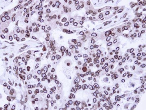
Immunohistochemical analysis of paraffin-embedded H441 Xenograft, using Lamin A + C antibody at 1:100 dilution.Application
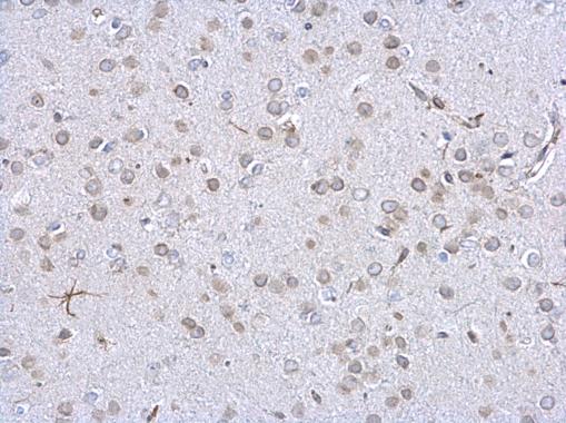
Lamin A + C antibody detects Lamin A + C protein at nuclear envelope on mouse fore brain by immunohistochemical analysis. Sample: Paraffin-embedded mouse fore brain. Lamin A + C antibody dilution: 1:500.Application
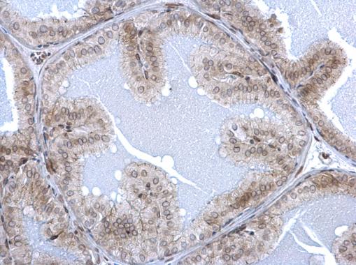
Lamin A + C antibody detects Lamin A + C protein at nuclear envelope on mouse prostate by immunohistochemical analysis. Sample: Paraffin-embedded mouse prostate. Lamin A + C antibody dilution: 1:500.Application
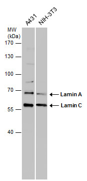
Various whole cell extracts (30 μg) were separated by 7.5% SDS-PAGE, and the membrane was blotted with Lamin A + C antibody diluted at 1:1000.| Product Name | Lamin A + C antibody |
|---|---|
| Antibody Type | Primary Antibodies |
| Product description | Rabbit Polyclonal antibody to Lamin A + CThe nuclear lamina consists of a two-dimensional matrix of proteins located next to the inner nuclear membrane. The lamin family of proteins make up the matrix and are highly conserved in evolution. During mitosis, the lamina matrix is reversibly disassembled as the lamin proteins are phosphorylated. Lamin proteins are thought to be involved in nuclear stability, chromatin structure and gene expression. Vertebrate lamins consist of two types, A and B. Through alternate splicing, this gene encodes three type A lamin isoforms. Mutations in this gene lead to several diseases: Emery-Dreifuss muscular dystrophy, familial partial lipodystrophy, limb girdle muscular dystrophy, dilated cardiomyopathy, Charcot-Marie-Tooth disease, and Hutchinson-Gilford progeria syndrome. [provided by RefSeq] |
| Immunogen | Recombinant protein encompassing a sequence within the center region of human Lamin A/C. The exact sequence is proprietary. |
| Clonality | Polyclonal |
|---|---|
| Isotype | IgG |
| Host Species | Rabbit |
| Tested Applications | IHC-PWB |
| *Optimal dilutions/concentrations should be determined by the researcher.
: |
|
| Species Reactivity | HumanMouse |
| Predicted Reactivity | Rat, Dog, Pig, Rhesus Monkey, Chimpanzee, BovineRat (98%), Dog (98%), Pig (98%), Rhesus Monkey (99%), Chimpanzee (100%), Bovine (98%) |
| Concentration | 1mg/ml (Please refer to the vial label for the specific concentration) |
| Purification | Affinity purified |
| Gene Synonyms | CDCD1 antibody CDDC antibody CMD1A antibody CMT2B1 antibody EMD2 antibody FPL antibody FPLD antibody HGPS antibody IDC antibody LDP1 antibody LFP antibody LGMD1B antibody LMN1 antibody LMNC antibody LMNL1 antibody PRO1 antibody LMNA antibod |
|---|---|
| Molecular Weight(MW) | 74 kDa (note) (5) |
| Cellular Localization | Nucleus |
Application
Lamin A + C antibody detects Lamin A + C protein by Western blot analysis. A. 30 μg NIH-3T3 whole cell lysate/extract 7.5 % SDS-PAGE Lamin A + C antibody dilution: 1:1000
Application
Sample (20 ug ) A: HeLa Nucleus 10% SDS PAGE diluted at 1:3000
Application
Immunohistochemical analysis of paraffin-embedded H441 Xenograft, using Lamin A + C antibody at 1:100 dilution.
Application
Lamin A + C antibody detects Lamin A + C protein at nuclear envelope on mouse fore brain by immunohistochemical analysis. Sample: Paraffin-embedded mouse fore brain. Lamin A + C antibody dilution: 1:500.
Application
Lamin A + C antibody detects Lamin A + C protein at nuclear envelope on mouse prostate by immunohistochemical analysis. Sample: Paraffin-embedded mouse prostate. Lamin A + C antibody dilution: 1:500.
Application
Various whole cell extracts (30 μg) were separated by 7.5% SDS-PAGE, and the membrane was blotted with Lamin A + C antibody diluted at 1:1000.| Positive Control | HeLa Nucleus , NIH-3T3 , A431 , NIH-3T3 |
|---|---|
| Application Notes | *Optimal dilutions/concentrations should be determined by the researcher.
: |
| Form | Liquid |
|---|---|
| Storage Instructions | Keep as concentrated solution. Aliquot and store at -20ºC or below. Avoid multiple freeze-thaw cycles. |
| Storage Buffer | 0.1M Tris, 0.1M Glycine, 10% Glycerol (pH7). 0.01% Thimerosal was added as a preservative. |


