Application
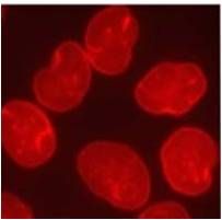
Immunofluorescent analysis of Lamin A/C (red) in HeLa cells.Application
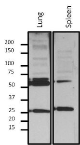
Western blot analysis of Lamin A/C in 10 μg of nuclear extract from fractionated mouse tissue lysate. Proteins were transferred to a PVDF membrane and blocked with 5% BSA/TBST for at least 1 hour. Membranes were then probed with Lamin A/C antibody [mab636] at a dilution of 1:500 overnight at 4°C on a rocking platform. Membranes were then washed in TBS-0.1%Tween 20 and probed with a HRP-conjugated secondary antibody. Membranes were washed and chemiluminescent detection was performed.| Product Name | Lamin A + C antibody |
|---|---|
| Antibody Type | Primary Antibodies |
| Product description | Mouse Monoclonal antibody [mab636] to Lamin A + CThe nuclear lamina consists of a two-dimensional matrix of proteins located next to the inner nuclear membrane. The lamin family of proteins make up the matrix and are highly conserved in evolution. During mitosis, the lamina matrix is reversibly disassembled as the lamin proteins are phosphorylated. Lamin proteins are thought to be involved in nuclear stability, chromatin structure and gene expression. Vertebrate lamins consist of two types, A and B. Alternative splicing results in multiple transcript variants. Mutations in this gene lead to several diseases: Emery-Dreifuss muscular dystrophy, familial partial lipodystrophy, limb girdle muscular dystrophy, dilated cardiomyopathy, Charcot-Marie-Tooth disease, and Hutchinson-Gilford progeria syndrome. [provided by RefSeq, Apr 2012] |
| Immunogen | Porcine lamin preparation. |
| Clonality | Monoclonal |
|---|---|
| Isotype | IgG2 |
| Clone Number | mab636 |
| Host Species | Mouse |
| Tested Applications | ICC/IFIHC-FWB |
| ICC/IF: 1:100: |
|
| Species Reactivity | HumanMousePigRatBovine |
| Concentration | 1mg/ml |
| Gene Synonyms | HGPS EMD2 FPLD lamin A/C LDP1 LMNA LMNL1 LMNC FPL LFP CDCD1 LGMD1B IDC CMT2B1 PRO1 FPLD2 A |
|---|---|
| Target | Lamin A + C |
Application
Immunofluorescent analysis of Lamin A/C (red) in HeLa cells.
Application
Western blot analysis of Lamin A/C in 10 μg of nuclear extract from fractionated mouse tissue lysate. Proteins were transferred to a PVDF membrane and blocked with 5% BSA/TBST for at least 1 hour. Membranes were then probed with Lamin A/C antibody [mab636] at a dilution of 1:500 overnight at 4°C on a rocking platform. Membranes were then washed in TBS-0.1%Tween 20 and probed with a HRP-conjugated secondary antibody. Membranes were washed and chemiluminescent detection was performed.| Application Notes | ICC/IF: 1:100: |
|---|
| Form | Liquid |
|---|---|
| Storage Instructions | -20° C, Avoid Freeze/Thaw Cycles |
| Storage Buffer | tissue culture supernatant containing 0.05 % sodium azide |


