Application
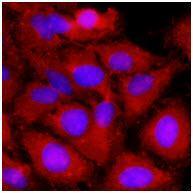
Immunofluorescence of human HeLa cells stained with monoclonal anti-human LTA antibody (1:500) with Texas Red (Red). Nucleus was stained by Hoechst 33342 (Blue).Application
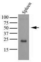
Tissue lysates of mouse spleen (35ug) were resolved by SDS-PAGE, transferred to NC membrane and probed with anti-human LTA (1:1000). Proteins were visualized using a goat anti-mouse secondary antibody conjugated to HRP and an ECL detection system.Application
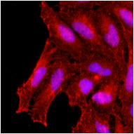
Immunofluorescence of human HeLa cells stained with monoclonal anti-human KCDT15 antibody (1:500, ) with Texas Red (Red). Nucleus was stained by Hoechst 33342 (Blue).Application
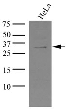
Cell lysates of HeLa (35ug) were resolved by SDS-PAGE, transferred to NC membrane and probed with anti-human KCTD15 (1:1000, ). Proteins were visualized using a goat anti-mouse secondary antibody conjugated to HRP and an ECL detection system.| Product Name | LTA antibody [AT15A3] |
|---|---|
| Antibody Type | Primary Antibodies |
| Product description | Mouse monoclonal antibody [AT15A3] to LTALymphotoxin-alpha (LT-alpha; LTA) is member of the TNF ligand superfamily, bind the same TNF receptor and mediate similar pleiotropic effect. LTA, located within the MHC III region of chromosome 6, show close linkage to the HLA class I (HLA-B) and class II (HLA-DR) genes. LTA is proinflammatory cytokine with important biological activity and immunomodulatory function and be known to influence a variety of cellular response. In particular, Polymorphism in the gene encoding LTA appears to contribute to infectious disease susceptibility and infection. |
| Immunogen | Recombinant human LTA (35-205aa) purified from E.coli |
| Clonality | Monoclonal |
|---|---|
| Isotype | IgG2 |
| Clone Number | AT15A3 |
| Host Species | Mouse |
| Tested Applications | ELISAICC/IFWB |
| Species Reactivity | HumanMouseRat |
| Concentration | 1mg/ml (Please refer to the vial label for the specific concentration) |
| Purification | Affinity purified |
| Gene Synonyms | OTTHUMP00000037613, TNF B, Tumor necrosis factor ligand supe, Lymphotoxin alpha, Tumor necrosis factor beta, TNF superfamily member 1, LTalpha, TNF superfamily member1, LTA, Coleya??s toxin, LT alpha, Natural killer cytotoxic factor, TNFB, Lymphotoxin alph |
|---|---|
| Target | LTA |
| Cellular Localization | The homotrimer is secreted. The heterotrimer is membrane-associated. |
| Light Chain Type | kappa |
Application
Immunofluorescence of human HeLa cells stained with monoclonal anti-human LTA antibody (1:500) with Texas Red (Red). Nucleus was stained by Hoechst 33342 (Blue).
Application
Tissue lysates of mouse spleen (35ug) were resolved by SDS-PAGE, transferred to NC membrane and probed with anti-human LTA (1:1000). Proteins were visualized using a goat anti-mouse secondary antibody conjugated to HRP and an ECL detection system.
Application
Immunofluorescence of human HeLa cells stained with monoclonal anti-human KCDT15 antibody (1:500, ) with Texas Red (Red). Nucleus was stained by Hoechst 33342 (Blue).
Application
Cell lysates of HeLa (35ug) were resolved by SDS-PAGE, transferred to NC membrane and probed with anti-human KCTD15 (1:1000, ). Proteins were visualized using a goat anti-mouse secondary antibody conjugated to HRP and an ECL detection system.| Form | Liquid |
|---|---|
| Storage Instructions | Can be stored at +4C. For long term storage, aliquot and store at -20C. Avoid repeated freezing and thawing cycles. |
| Storage Buffer | In Phosphate-Buffered Saline (pH 7.4) with 0.1% Sodium Azide |


