Application
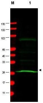
Western blot using GeneTex Mab anti-MAD2L1 antibody shows detection of a band at ~24 kDa (arrowhead) corresponding to MAD2L1 present in a HeLa whole cell lysate (lane 1). Approximately 75 µg of lysate was separated by 4-20% TG SDS-PAGE. After blocking, the membrane was probed overnight at 4°C with the primary antibody diluted to 1:200. The membrane was washed and reacted with a 1:5,000 dilution of IRDye™800 conjugated Sh-a-Mouse IgG [H&L] for 45 min at room temperature (800 nm channel, green). Molecular weight estimation was made by comparison to prestained MW markers in lane M (700 nm channel, red). IRDye™800 fluorescence image was captured using the Odyssey® Infrared Imaging System developed by LI-COR. IRDye is a trademark of LI-COR, Inc.| Product Name | MAD2L1 antibody [17D10] |
|---|---|
| Antibody Type | Primary Antibodies |
| Product description | Mouse Monoclonal antibody [17D10] to MAD2L1MAD2L1 (also called mitotic spindle assembly checkpoint protein, MAD2A, MAD2-like 1 and HsMAD2) is a component of the mitotic spindle assembly checkpoint monitors the process of kinetochore-spindle attachment and delays the onset of anaphase when this process is not complete. MAD2L1 inhibits the activity of the anaphase-promoting complex by sequestering CDC20 until all chromosomes are aligned at the metaphase plate. MAD2L1 is related to the MAD2L2 gene located on chromosome 1. This protein has a nuclear localization. |
| Immunogen | This protein A purified monoclonal antibody was produced by repeated immunizations with full-length recombinant human MAD2L1 protein. |
| Clonality | Monoclonal |
|---|---|
| Isotype | IgG1 |
| Clone Number | 17D10 |
| Host Species | Mouse |
| Tested Applications | ELISAIPWB |
| *Optimal dilutions/concentrations should be determined by the researcher. | |
| Species Reactivity | Human |
| Concentration | 1mg/ml |
| Purification | Protein A |
| Gene Synonyms | HSMAD2 , MAD2 |
|---|---|
| Target | MAD2L1 |
| Cellular Localization | Nuclear |
Application
Western blot using GeneTex Mab anti-MAD2L1 antibody shows detection of a band at ~24 kDa (arrowhead) corresponding to MAD2L1 present in a HeLa whole cell lysate (lane 1). Approximately 75 µg of lysate was separated by 4-20% TG SDS-PAGE. After blocking, the membrane was probed overnight at 4°C with the primary antibody diluted to 1:200. The membrane was washed and reacted with a 1:5,000 dilution of IRDye™800 conjugated Sh-a-Mouse IgG [H&L] for 45 min at room temperature (800 nm channel, green). Molecular weight estimation was made by comparison to prestained MW markers in lane M (700 nm channel, red). IRDye™800 fluorescence image was captured using the Odyssey® Infrared Imaging System developed by LI-COR. IRDye is a trademark of LI-COR, Inc.| Application Notes | *Optimal dilutions/concentrations should be determined by the researcher. |
|---|
| Form | Liquid |
|---|---|
| Storage Instructions | Store vial at -20° C prior to opening. Aliquot contents and freeze at -20° C or below for extended storage. Avoid cycles of freezing and thawing. |
| Storage Buffer | 0.02 M Potassium Phosphate, 0.15 M Sodium Chloride, pH 7.2 and 0.01% (w/v) Sodium Azide |


