Application
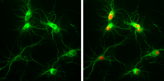
MAP2 antibody [HM-2] detects MAP2 protein at by immunofluorescent analysis. Sample: Rat E18 primary cortical neuron, DIV 8. Cells were fixed in 4% paraformaldehyde at RT for 15 min. Green: MAP2 protein stained by MAP2 antibody [HM-2] diluted at 1:250. Red: NeuN, stained by NeuN antibody diluted at 1:250.Application
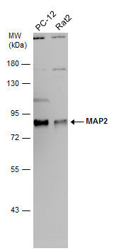
Various whole cell extracts (30 µg) were separated by 7.5% SDS-PAGE, and the membrane was blotted with MAP2 antibody [HM-2] diluted at 1:2000. The HRP-conjugated anti-mouse IgG antibody was used to detect the primary antibody, and the signal was developed with Trident ECL plus-Enhanced.Application
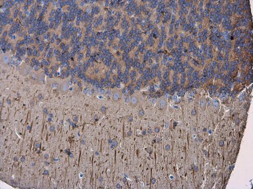
MAP2 antibody [HM-2] detects MAP2 protein at cytoplasm in mouse brain by immunohistochemical analysis. Sample: Paraffin-embedded mouse brain. MAP2 antibody [HM-2] diluted at 1:500.Application
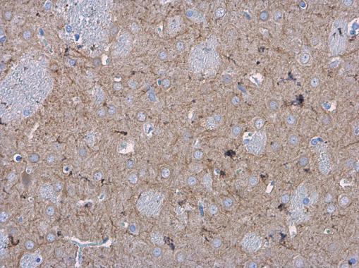
MAP2 antibody [HM-2] detects MAP2 protein at cytoplasm in rat brain by immunohistochemical analysis. Sample: Paraffin-embedded rat brain. MAP2 antibody [HM-2] diluted at 1:500.Application
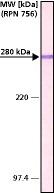
Rat brain extract (adult and newborn) was separated on SDS-PAGE and probed with Monoclonal PA To MAP2 (MO) CL:HM-2 . The antibody was developed with AlkPhos APA Mouse Fab ads HIgG and a NBTBCIP substrate. Antibody concentration: 1 μg/mL.Application
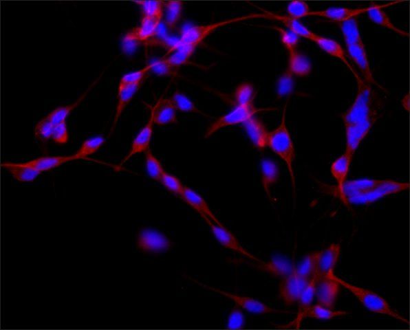
ICC/IF analysis of B35 cells using MAP2 antibody [HM-2] at a concentration of 2 μg/ml (red) and DAPI (blue).| Product Name | MAP2 antibody [HM-2] |
|---|---|
| Antibody Type | Primary Antibodies |
| Product description | Mouse monoclonal [HM - 2] to MAP2MAP2 is the major microtubule associated protein of brain tissue. There are three forms of MAP2; two are similarily sized with apparent molecular weights of 280 kDa (MAP2a and MAP2b) and the third with a lower molecular weight of 70 kDa (MAP2c). In the newborn rat brain, MAP2b and MAP2c are present, while MAP2a is absent. Between postnatal days 10 and 20, MAP2a appears. At the same time, the level of MAP2c drops by 10-fold. This change happens during the period when dendrite growth is completed and when neurons have reached their mature morphology. MAP2 is degraded by a Cathepsin D-like protease in the brain of aged rats. There is some indication that MAP2 is expressed at higher levels in some types of neurons than in other types. MAP2 is known to promote microtubule assembly and to form side-arms on microtubules. It also interacts with neurofilaments, actin, and other elements of the cytoskeleton. |
| Immunogen | Rat brain microtubule associated proteins (MAPs). |
| Clonality | Monoclonal |
|---|---|
| Isotype | IgG1 |
| Clone Number | HM-2 |
| Host Species | Mouse |
| Tested Applications | ICC/IFIHCIHC-FIHC-PWB |
| For ICC/IF: 1:100-1:500. For IHC (suitable): Use at an assay dependent dilution. For IHC-P: 1:100-1:500. For WB: 1:500-1:2000. Optimal dilutions/concentrations should be determined by the researcher. | |
| Species Reactivity | HumanMouseQuailRat |
| Concentration | 1mg/ml (Please refer to the vial label for the specific concentration) |
| Purification | Protein A |
| Gene Synonyms | MAP2R, Mtap2 |
|---|---|
| Target | MAP2 |
| Tissue Specificity | Monoclonal Anti-MAP2 does not react with tubulin of other microtubule associated proteins. |
| Cellular Localization | Cytoplasmic |
Application
MAP2 antibody [HM-2] detects MAP2 protein at by immunofluorescent analysis. Sample: Rat E18 primary cortical neuron, DIV 8. Cells were fixed in 4% paraformaldehyde at RT for 15 min. Green: MAP2 protein stained by MAP2 antibody [HM-2] diluted at 1:250. Red: NeuN, stained by NeuN antibody diluted at 1:250.
Application
Various whole cell extracts (30 µg) were separated by 7.5% SDS-PAGE, and the membrane was blotted with MAP2 antibody [HM-2] diluted at 1:2000. The HRP-conjugated anti-mouse IgG antibody was used to detect the primary antibody, and the signal was developed with Trident ECL plus-Enhanced.
Application
MAP2 antibody [HM-2] detects MAP2 protein at cytoplasm in mouse brain by immunohistochemical analysis. Sample: Paraffin-embedded mouse brain. MAP2 antibody [HM-2] diluted at 1:500.
Application
MAP2 antibody [HM-2] detects MAP2 protein at cytoplasm in rat brain by immunohistochemical analysis. Sample: Paraffin-embedded rat brain. MAP2 antibody [HM-2] diluted at 1:500.
Application
Rat brain extract (adult and newborn) was separated on SDS-PAGE and probed with Monoclonal PA To MAP2 (MO) CL:HM-2 . The antibody was developed with AlkPhos APA Mouse Fab ads HIgG and a NBTBCIP substrate. Antibody concentration: 1 μg/mL.
Application
ICC/IF analysis of B35 cells using MAP2 antibody [HM-2] at a concentration of 2 μg/ml (red) and DAPI (blue).| Application Notes | For ICC/IF: 1:100-1:500. For IHC (suitable): Use at an assay dependent dilution. For IHC-P: 1:100-1:500. For WB: 1:500-1:2000. Optimal dilutions/concentrations should be determined by the researcher. |
|---|
| Form | Liquid |
|---|---|
| Storage Instructions | Aliquot and store at -20°C or -80°C. Avoid repeated freeze / thaw cycles. |
| Storage Buffer | 0.01 M phosphate-buffered saline, pH 7.4, containing 15 mM sodium azide |


