WB
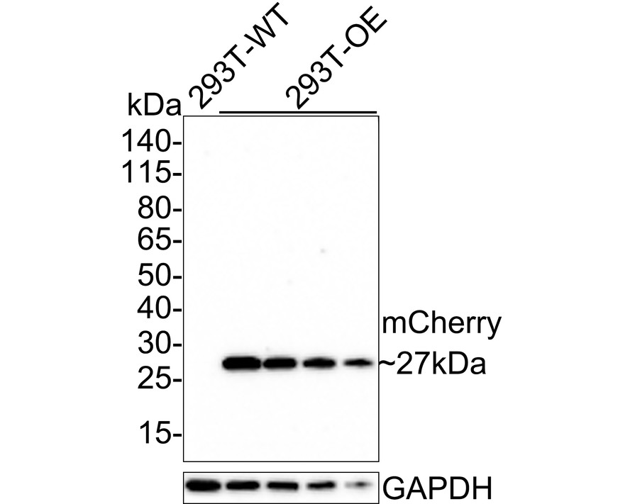
Western blot analysis of mCherry on different lysates with Mouse anti-mCherry antibody at 1/2,000 dilution. Lane 1: 293T-WT cell lysate (5 µg/Lane), Lane 2: 293T-OE-mCherry cell lysate (5 µg/Lane), Lane 3: 293T-OE-mCherry cell lysate (2.5 µg/Lane), Lane 4: 293T-OE-mCherry cell lysate (1.25 µg/Lane), Lane 5: 293T-OE-mCherry cell lysate (0.625 µg/Lane), Exposure time: 30 seconds; 4-20% SDS-PAGE gel. Proteins were transferred to a PVDF membrane and blocked with 5% NFDM/TBST for 1 hour at room temperature. The primary antibody at 1/2,000 dilution was used in 5% NFDM/TBST at room temperature for 2 hours. Goat Anti-Mouse IgG - HRP Secondary Antibody at 1/150,000 dilution was used for 1 hour at room temperature.WB
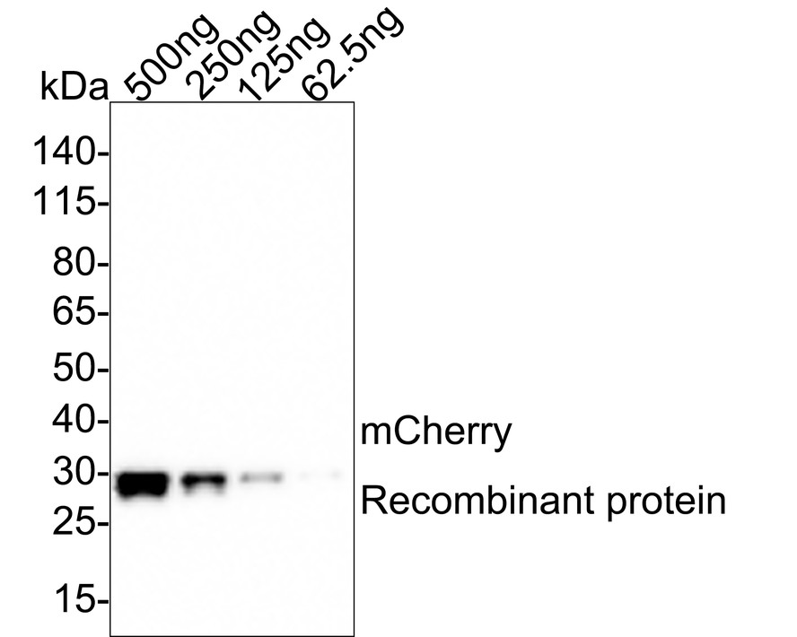
Western blot analysis of mCherry on mCherry recombinant protein with Mouse anti-mCherry antibody at 1/1,000 dilution. Lane 1: mCherry recombinant protein, 500ng/Lane, Lane 2: mCherry recombinant protein, 250ng/Lane, Lane 3: mCherry recombinant protein, 125ng/Lane, Lane 4: mCherry recombinant protein, 62.5ng/Lane, Exposure time: 30 seconds; 4-20% SDS-PAGE gel. Proteins were transferred to a PVDF membrane and blocked with 5% NFDM/TBST for 1 hour at room temperature. The primary antibody at 1/1,000 dilution was used in 5% NFDM/TBST at room temperature for 2 hours. Goat Anti-Mouse IgG - HRP Secondary Antibody at 1/150,000 dilution was used for 1 hour at room temperature.ICC/IF
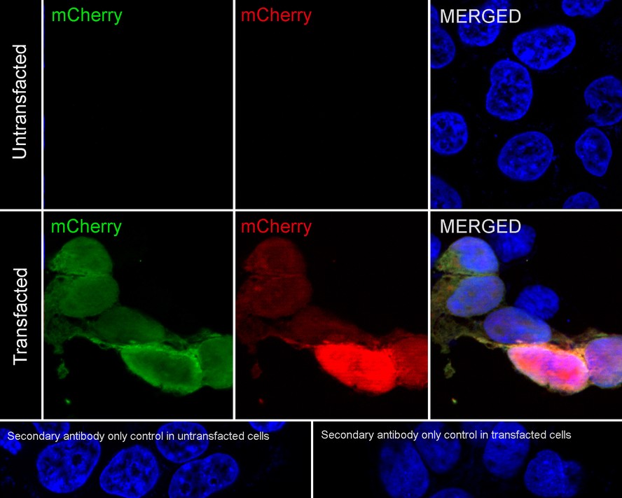
Immunocytochemistry analysis of 293T transfected with or without mCherry cells labeling mCherry with Mouse anti-mCherry antibody at 1/2,000 dilution. Cells were fixed in 4% paraformaldehyde for 20 minutes at room temperature, permeabilized with 0.1% Triton X-100 in PBS for 5 minutes at room temperature, then blocked with 1% BSA in 10% negative goat serum for 1 hour at room temperature. Cells were then incubated with Mouse anti-mCherry antibody at 1/2,000 dilution in 1% BSA in PBST overnight at 4 ℃. Goat Anti-Mouse IgG H&L (488) was used as the secondary antibody at 1/1,000 dilution. PBS instead of the primary antibody was used as the secondary antibody only control. Nuclear DNA was labelled in blue with DAPI. As expected, green antibody staining is only visible in cells expressing mCherry, and green and red signals can be superimposed.| Product Name | mCherry Mouse Monoclonal Antibody |
|---|---|
| Antibody Type | Primary Antibodies |
| Product description | Mouse Monoclonal antibody [GT857] to mCherry |
| Immunogen | Recombinant protein within mCherry aa 1-236 / 236. |
| Clonality | Monoclonal |
|---|---|
| Isotype | IgG2b |
| Host Species | Mouse |
| Tested Applications | ICC/IFWB |
| WB:1:2000-1:5000 ICC/IF:1:2000 |
|
| Species Reactivity | All |
| Concentration | 1mg/ml |
| Purification | Protein A |
| Molecular Weight(MW) | 27kDa |
|---|---|
| Function | mCherry is a member of the mFruits family of monomeric red fluorescent proteins (mRFPs). As a RFP, mCherry was derived from DsRed of Discosoma sea anemones unlike green fluorescent proteins (GFPs) which are often derived from Aequoera victoria jellyfish.Fluorescent proteins are used to tag components in the cell, so they can be studied using fluorescence spectroscopy and fluorescence microscopy. mCherry absorbs light between 540-590 nm and emits light in the range of 550-650 nm. mCherry belongs to the group of fluorescent protein chromophores used as instruments to visualize genes and analyze their functions in experiments. Genome editing has been improved greatly through the precise insertion of these fluorescent protein tags into the genetic material of many diverse organisms. Most comparisons between the brightness and photostability of different fluorescent proteins have been made in vitro, removed from biological variables that affect protein performance in cells or organisms. It is hard to perfectly simulate cellular environments in vitro, and the difference in environment could have an effect on the brightness and photostability. |
WB
Western blot analysis of mCherry on different lysates with Mouse anti-mCherry antibody at 1/2,000 dilution. Lane 1: 293T-WT cell lysate (5 µg/Lane), Lane 2: 293T-OE-mCherry cell lysate (5 µg/Lane), Lane 3: 293T-OE-mCherry cell lysate (2.5 µg/Lane), Lane 4: 293T-OE-mCherry cell lysate (1.25 µg/Lane), Lane 5: 293T-OE-mCherry cell lysate (0.625 µg/Lane), Exposure time: 30 seconds; 4-20% SDS-PAGE gel. Proteins were transferred to a PVDF membrane and blocked with 5% NFDM/TBST for 1 hour at room temperature. The primary antibody at 1/2,000 dilution was used in 5% NFDM/TBST at room temperature for 2 hours. Goat Anti-Mouse IgG - HRP Secondary Antibody at 1/150,000 dilution was used for 1 hour at room temperature.
WB
Western blot analysis of mCherry on mCherry recombinant protein with Mouse anti-mCherry antibody at 1/1,000 dilution. Lane 1: mCherry recombinant protein, 500ng/Lane, Lane 2: mCherry recombinant protein, 250ng/Lane, Lane 3: mCherry recombinant protein, 125ng/Lane, Lane 4: mCherry recombinant protein, 62.5ng/Lane, Exposure time: 30 seconds; 4-20% SDS-PAGE gel. Proteins were transferred to a PVDF membrane and blocked with 5% NFDM/TBST for 1 hour at room temperature. The primary antibody at 1/1,000 dilution was used in 5% NFDM/TBST at room temperature for 2 hours. Goat Anti-Mouse IgG - HRP Secondary Antibody at 1/150,000 dilution was used for 1 hour at room temperature.
ICC/IF
Immunocytochemistry analysis of 293T transfected with or without mCherry cells labeling mCherry with Mouse anti-mCherry antibody at 1/2,000 dilution. Cells were fixed in 4% paraformaldehyde for 20 minutes at room temperature, permeabilized with 0.1% Triton X-100 in PBS for 5 minutes at room temperature, then blocked with 1% BSA in 10% negative goat serum for 1 hour at room temperature. Cells were then incubated with Mouse anti-mCherry antibody at 1/2,000 dilution in 1% BSA in PBST overnight at 4 ℃. Goat Anti-Mouse IgG H&L (488) was used as the secondary antibody at 1/1,000 dilution. PBS instead of the primary antibody was used as the secondary antibody only control. Nuclear DNA was labelled in blue with DAPI. As expected, green antibody staining is only visible in cells expressing mCherry, and green and red signals can be superimposed.| Application Notes | WB:1:2000-1:5000 ICC/IF:1:2000 |
|---|
| Form | Liquid |
|---|---|
| Storage Instructions | Store at +4℃ short term (1-2 weeks). It is recommended to aliquot into single-use upon delivery. Store at -20℃ long term. |
| Storage Buffer | PBS (pH7.4), 0.1% BSA, 40% Glycerol. Preservative: 0.05% Sodium Azide. |


