Application
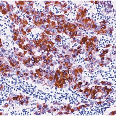
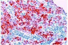
Formalin fixed paraffin embedded human melanoma stained with Melanoma antibody .Application
IHC Image using - Detection of Melanoma by IHC in Human Melanoma| Product Name | Melanoma antibody [HMB45] |
|---|---|
| Antibody Type | Primary Antibodies |
| Product description | Mouse monoclonal [HMB45] to Melanoma |
| Immunogen | Extract of pigmented melanoma metastases from lymph nodes. |
| Clonality | Monoclonal |
|---|---|
| Isotype | IgG1 |
| Clone Number | HMB45 |
| Host Species | Mouse |
| Tested Applications | IHCIHC-FIHC-P |
| IHC: This antibody may be diluted to a titer of 1:10-1:50 in an ABC method. Staining Protocol: We suggest an incubation period of 30-60 minutes at room temperature. However, depending upon the fixation conditions and the staining system employed, optimal incubation conditions and antibody dilutions should be determined by the user. Enzymatic antigen retrieval may increase staining intensity when using formalin fixed paraffin embedded tissue. | |
| Species Reactivity | Human |
| Concentration | 1mg/ml |
| Purification | Unpurified |
| Gene Synonyms | CMM1, DNS, FAMMM, MLM |
|---|---|
| Target | Melanoma |
| Tissue Specificity | This antibody reacts with a neuraminidase sensitive oligosaccharide side chain of a glycoconjugate present in immature melanosomes. It reacts with junctional and blue nevus cells. Non-melanocytic cells are negative. |
| Cellular Localization | Cytoplasmic |
| Light Chain Type | kappa |
Application
Formalin fixed paraffin embedded human melanoma stained with Melanoma antibody .
Application
IHC Image using - Detection of Melanoma by IHC in Human Melanoma| Application Notes | IHC: This antibody may be diluted to a titer of 1:10-1:50 in an ABC method. Staining Protocol: We suggest an incubation period of 30-60 minutes at room temperature. However, depending upon the fixation conditions and the staining system employed, optimal incubation conditions and antibody dilutions should be determined by the user. Enzymatic antigen retrieval may increase staining intensity when using formalin fixed paraffin embedded tissue. |
|---|
| Form | Liquid |
|---|---|
| Storage Instructions | Keep as concentrated solution, aliquot and store at 4°C. |
| Storage Buffer | Tissue culture supernatant with PBS, 1% BSA and 0.05% sodium azide |


