Application
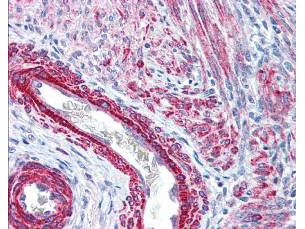
Myosin Light chain (phospho Ser20) antibody was used at 2.5 µg/ml to detect signal in a variety of tissues including multi-human, multi-brain and multi-cancer slides. This image shows strong staining of both vascular and myometrial smooth muscle cells of the uterus. Tissue was formalin-fixed and paraffin embedded. The image shows localization of the antibody as the precipitated red signal, with a hematoxylin purple nuclear counterstain.Application
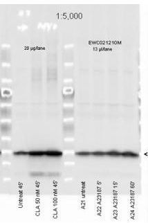
Myosin Light chain (phospho Ser20) antibody was used at a 1:5000 dilution to detect myosin light chain by Western blot. Either 13 or 20 ul of a mouse cardiac myocyte lysate was loaded on a 4-20% Criterion gel for SDS-PAGE. Samples were either mock-treated or CLA-treated, as indicated. After washing, a 1:5,000 dilution of HRP conjugated Gt-a-Rabbit IgG preceded color development using Amersham's substrate systemApplication
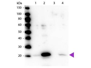
Western blot of Myosin Light chain (phospho Ser20) antibody . Lane 1: Regulatory Light Chain Non-Phospho recombinant protein. Lane 2: Regulatory Light Chain Phospho recombinant protein. Lane 3: Smooth Muscle Non-Phospho recombinant protein. Lane 4: Smooth Muscle Phospho recombinant protein. Load: 50 ng per lane. Primary antibody at 1:1,000 overnight at 4°C. Secondary antibody: Peroxidase rabbit secondary antibody at 1:40,000 for 60 min at RT. Blocking for 30 min at RT. Predicted/Observed size: 20 kDa.| Product Name | Myosin Light chain (phospho Ser20) antibody |
|---|---|
| Antibody Type | Primary Antibodies |
| Product description | Rabbit Polyclonal antibody to Myosin Light chain (phospho Ser20)Myosin is the major component of thick muscle filaments, and is a long asymmetric molecule containing a globular head and a long tail. The molecule consists of two heavy chains each ~200,000 daltons, and four light chains each ~16,000 - 21,000 daltons. Activation of smooth and cardiac muscle primarily involves pathways that increase calcium levels and myosin phosphorylation, resulting in contraction. Myosin light chain phosphatase acts to regulate muscle contraction by dephosphorylating activated myosin light chain. This antibody is specific for the phosphorylated form of myosin light chain. The selected peptide sequence used to generate the polyclonal antibody is located near the amino terminal end of the polypeptide corresponding to the smooth/non-muscle form of myosin regulatory light chain found in cardiac myocytes in addition to smooth and non-muscle cells. This sequence differs from that of the sarcomeric/cardiac form of myosin regulatory light chain that has a different sequence around the phosphorylation site. Human and mouse have almost identical sequences. In human the phosphorylation site is pS19, while in mouse the site maps to pS20. |
| Immunogen | Human Myosin Light Chain phospho peptide corresponding to a region near the amino terminus of the human smooth/non-muscle form of myosin regulatory light chain conjugated to KLH - KKRPQRAT(pS)NVFAMFD (aa 12-27). |
| Modification | p-Ser20 |
| Clonality | Polyclonal |
|---|---|
| Isotype | IgG |
| Host Species | Rabbit |
| Tested Applications | ELISAICC/IFIHC-PIPWB |
| *Optimal dilutions/concentrations should be determined by the researcher.
: |
|
| Species Reactivity | HumanMouse |
| Concentration | 1mg/ml |
| Purification | Affinity purified |
| Gene Synonyms | MRLC3 MLCB MYL2B MRCL3 MYL12A |
|---|---|
| Target | Myosin Light chain |
| Cellular Localization | Cytoplasmic |
Application
Myosin Light chain (phospho Ser20) antibody was used at 2.5 µg/ml to detect signal in a variety of tissues including multi-human, multi-brain and multi-cancer slides. This image shows strong staining of both vascular and myometrial smooth muscle cells of the uterus. Tissue was formalin-fixed and paraffin embedded. The image shows localization of the antibody as the precipitated red signal, with a hematoxylin purple nuclear counterstain.
Application
Myosin Light chain (phospho Ser20) antibody was used at a 1:5000 dilution to detect myosin light chain by Western blot. Either 13 or 20 ul of a mouse cardiac myocyte lysate was loaded on a 4-20% Criterion gel for SDS-PAGE. Samples were either mock-treated or CLA-treated, as indicated. After washing, a 1:5,000 dilution of HRP conjugated Gt-a-Rabbit IgG preceded color development using Amersham's substrate system
Application
Western blot of Myosin Light chain (phospho Ser20) antibody . Lane 1: Regulatory Light Chain Non-Phospho recombinant protein. Lane 2: Regulatory Light Chain Phospho recombinant protein. Lane 3: Smooth Muscle Non-Phospho recombinant protein. Lane 4: Smooth Muscle Phospho recombinant protein. Load: 50 ng per lane. Primary antibody at 1:1,000 overnight at 4°C. Secondary antibody: Peroxidase rabbit secondary antibody at 1:40,000 for 60 min at RT. Blocking for 30 min at RT. Predicted/Observed size: 20 kDa.| Positive Control | Cardiac myocytes |
|---|---|
| Application Notes | *Optimal dilutions/concentrations should be determined by the researcher.
: |
| Form | Liquid |
|---|---|
| Storage Instructions | Store vial at -20° C prior to opening. Aliquot contents and freeze at -20° C or below for extended storage. Avoid cycles of freezing and thawing. |
| Storage Buffer | 0.02 M Potassium Phosphate, 0.15 M Sodium Chloride, pH 7.2 and 0.01% (w/v) Sodium Azide |


