Application
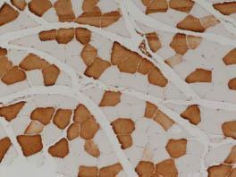
Slow skeletal fibers were immunostained (brown) using monoclonal anti-slow myosin antibody [NOQ7.5.4] at a dilution of 1:6000Application
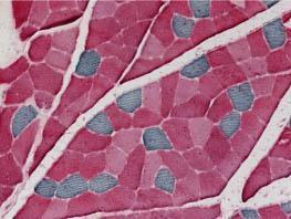
Double staining of skeletal muscle fibers using monoclonal anti-slow myosin antibody [NOQ7.5.4] at a dilution of 1:6000 (blue-gray) to label type I fibers and anti-fast myosin antibody (pink) to label type II fibersApplication
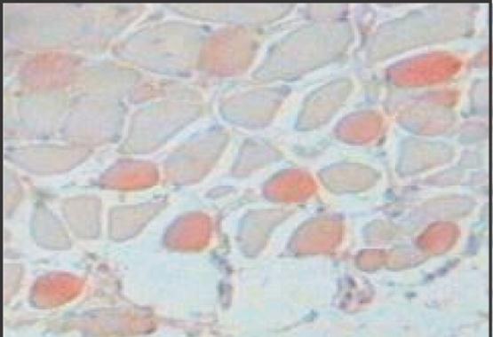
Staining of formalin-fixed, paraffin-embedded section of rabbit tongue using Monoclonal Anti-Myosin (Skeletal, Slow) . Antibody dilution 1:4,000. Specific staining of slow muscle myosin.Application
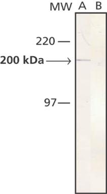
Whole cell extract of rabbit tongue was separated on SDS-PAGE and blotted with Monoclonal Anti-Myosin (Skeletal, Slow) . The antibody was developed with Goat Anti-Mouse IgG (Fab), Alkaline Phosphatase Conjugate and NBT/BCIP substrate. Lane A: Antibody dilution 1:5,000. Lane B: Negative control (only secondary antibody)| Product Name | Myosin skeletal slow antibody |
|---|---|
| Antibody Type | Primary Antibodies |
| Product description | Mouse monoclonal [NOQ7.5.4D] to Slow skeletal MyosinMyosin is a 480 kDa protein known to interact with actin in muscle and in non muscle cells. It contains two identical heavy chains (200 kDa each) and four light chains (15-26 kDa). Myosin molecules consist of two major regions: tail (rod) and heads; they aggregate into filaments through the tail region and interact with actin and with ATP through the head region. Multiple forms of myosin heavy chains exist for each muscle type: skeletal, cardiac, smooth and in non muscle. Myosin isoforms exist in different types of skeletal muscle, depending on the physiological function of the muscle. Mammalian muscle fibers are classified primarily into slow (I), fast-red (IIa) and fast-white (IIb) major types.4,9,10 Changes in the speed of muscle contraction brought about by neural influences result from changes in the pattern of expression of myosin and other myofibrillar genes. Myosin heavy chains are encoded by a multigene family which is developmentally regulated. During differentiation of future muscles, fibers sequentially express heavy chain genes for embryonic, neonatal and adult myosin. Developing muscles express two isoforms of myosin, the embryonic and fetal (neonatal) forms, which are also encoded for by distinct heavy chain genes. The embryonic and fetal myosins have a high calcium-activated ATPase activity at alkaline pH; fibers that contain these myosins are therefore likely to appear similar, histochemically, to mature Type II fibers, even though the latter contain qualitatively distinct mature fast isomyosins. In the neonate, the principal myosin in developing fast and slow muscles is fetal myosin. During the first few weeks of life, fetal myosin is replaced by slow myosin in slow muscle and by fast myosins in fast muscle. There are three adult forms of skeletal myosin corresponding to the three types of muscle fibers. The heavy chains of these myosins are distinct, and are encoded by different heavy chain genes. Muscle fiber type classification for type I and type II fibers has long been applied to the histological study of both normal and pathological human skeletal muscle. Studies of myosin isoforms expression in developing skeletal muscle also provide information about the control of gene expression in different muscle types by neural, hormonal and other factors. The advent of myosin immunocytochemistry, using antibodies to specific myosin heavy chains, has made it possible to trace the developmental history of the complex paths of myosin changes at the cellular level. Antibodies are also important because of the clues they may give concerning the pathogenesis of abnormalities in diseased muscle fibers. Monoclonal antibodies that recognize epitopes peculiar to a particular developmental or mature isomyosin may be used to identify these isomyosins and thus to characterize the temporal pattern of their expression in different fibers during myogenesis. |
| Immunogen | Full length native protein (purified) (Human). |
| Clonality | Monoclonal |
|---|---|
| Isotype | IgG1 |
| Clone Number | NOQ7.5.4D |
| Host Species | Mouse |
| Tested Applications | EMELISAIHCIHC-PRIAWB |
| For EM: Use at an assay dependent dilution. For ELISA: Use at an assay dependent dilution. For IHC-P: Use at a dilution of 1:4,000. Perform enzymatic antigen retrieval before commencing with IHC staining protocol. Detection of slow myosin in formalin-fixed,paraffin-embedded tissues, is greatly enhanced byproteolytic digestion of the preparation. For RIA: Use at an assay dependent dilution. For WB: Use at an assay dependent dilution. Predicted molecular weight: 15-26 kDa. Using immunohistochemical staining, the antibody reacts with human adult type I skeletal muscle fibers and with cardiac muscle cells. It also reacts faintly with adult human C-Fibers, co-expressing myosin heavy chain I and IIa in variable ratios. Optimal dilutions/concentrations should be determined by the end user.: |
|
| Species Reactivity | HumanMouseDogPigRatCatGoatGuinea PigHamsterRabbitSheepBovine |
| Concentration | 1mg/ml |
| Purification | Affinity purified |
| Target | Myosin skeletal slow |
|---|---|
| Tissue Specificity | Reacts heavy meromyosin portion of human adult skeletal muscle slow myosin. Does not react with the fast myosin heavy chain nor with embryonic or neonatal rat skeletal myosins, nor with cat fetal/embryonic skeletal myosin. Also reacts with human adult type I skeletal muscle fibers and with cardiac muscle cells. No reactivity detected with human or rat adult smooth muscle or with non-muscle tissues. |
| Cellular Localization | Cytoplasmic |
Application
Slow skeletal fibers were immunostained (brown) using monoclonal anti-slow myosin antibody [NOQ7.5.4] at a dilution of 1:6000
Application
Double staining of skeletal muscle fibers using monoclonal anti-slow myosin antibody [NOQ7.5.4] at a dilution of 1:6000 (blue-gray) to label type I fibers and anti-fast myosin antibody (pink) to label type II fibers
Application
Staining of formalin-fixed, paraffin-embedded section of rabbit tongue using Monoclonal Anti-Myosin (Skeletal, Slow) . Antibody dilution 1:4,000. Specific staining of slow muscle myosin.
Application
Whole cell extract of rabbit tongue was separated on SDS-PAGE and blotted with Monoclonal Anti-Myosin (Skeletal, Slow) . The antibody was developed with Goat Anti-Mouse IgG (Fab), Alkaline Phosphatase Conjugate and NBT/BCIP substrate. Lane A: Antibody dilution 1:5,000. Lane B: Negative control (only secondary antibody)| Application Notes | For EM: Use at an assay dependent dilution. For ELISA: Use at an assay dependent dilution. For IHC-P: Use at a dilution of 1:4,000. Perform enzymatic antigen retrieval before commencing with IHC staining protocol. Detection of slow myosin in formalin-fixed,paraffin-embedded tissues, is greatly enhanced byproteolytic digestion of the preparation. For RIA: Use at an assay dependent dilution. For WB: Use at an assay dependent dilution. Predicted molecular weight: 15-26 kDa. Using immunohistochemical staining, the antibody reacts with human adult type I skeletal muscle fibers and with cardiac muscle cells. It also reacts faintly with adult human C-Fibers, co-expressing myosin heavy chain I and IIa in variable ratios. Optimal dilutions/concentrations should be determined by the end user.: |
|---|
| Form | Liquid |
|---|---|
| Storage Instructions | Aliquot and store at -20°C or -80°C. Avoid repeated freeze / thaw cycles. |
| Storage Buffer | Ascitic fluid containing 15mM sodium azide |


