Application
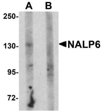
Western blot analysis of NALP5 in mouse brain tissue lysate with NALP5 antibody at 1 ug/ml in (A) the absence and (B) the presence of blocking peptide.Application
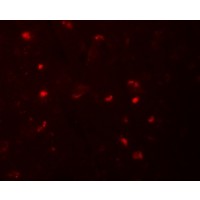
Immunofluorescence of NALP6 in human brain tissue with NALP6 antibody at 20 μg/mL.IHC
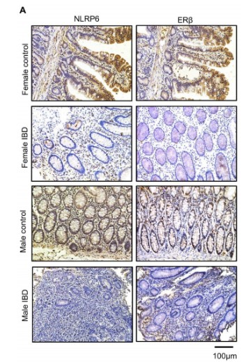
(A) Immunohistochemistry analysis of NLRP6 and ERβ in different intestinal tissue sections. (Scale bars, 100 µm).WB
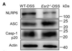
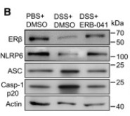
(A) NLRP6, ASC, and Casp-1 p20 expression of mouse colon homogenate.WB
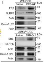
(I) ERβ, NLRP6, ASC, and Casp-1 p20 expression of mice colon homogenate. See also Figure S1 and Data S1.WB
(B) Immunoblotting of treated cell lysates. Data are representative of three independent experiments. ∗p < 0.05, ∗∗p < 0.01, by two-way ANOVA with Tukey’s post hoc analysis. Normal NCM-460 cells were stimulated with the selective ERβ agonist (ERB-041, 1 μM) and ERα agonist (PPT, 1 μM) for 48 h.| Product Name | NALP6 antibody |
|---|---|
| Antibody Type | Primary Antibodies |
| Product description | Rabbit polyclonal antibody to NALP6NALP proteins include the apoptosis regulator APAF1 (apoptotic protease activating factor 1) and mammalian NOD-LRR proteins and are thought to be involved in inflammation and reproduction. NALP6, also known as PYPAF5, is highly expressed in granulocytes and T cells and co-localizes with the PYRIN-CARD protein ASC. Co-expression of NALP6 with ASC results in a synergistic activation of NF-kB and activated caspase-1-dependent cytokine processing, suggesting that NALP6 functions in immune cells to coordinate the transduction of pro-inflammatory signals. |
| Clonality | Polyclonal |
|---|---|
| Host Species | Rabbit |
| Tested Applications | ELISAICC/IFWB |
| ICC/IF:1:200~1:500 WB:1:500~1:2000 Notes:Optimal dilutions/concentrations should be determined by the researcher. |
|
| Species Reactivity | Human |
| Concentration | 1mg/ml |
| Purification | Affinity purified |
| Gene Synonyms | NLR family pyrin domain containing6 PAN3 NLRP6 NLR family pyrin domain containing 6 CLR11.4 PYPAF5 NALP6 PYRINcontaining APAF1like protein5 PYRIN-containing APAF1-like protein 5 PYRINcontaining APAF1like protein 5 |
|---|---|
| Target | NALP6 |
| Tissue Specificity | This antibody is specific for NALP6 |
Application
Western blot analysis of NALP5 in mouse brain tissue lysate with NALP5 antibody at 1 ug/ml in (A) the absence and (B) the presence of blocking peptide.
Application
Immunofluorescence of NALP6 in human brain tissue with NALP6 antibody at 20 μg/mL.
IHC
(A) Immunohistochemistry analysis of NLRP6 and ERβ in different intestinal tissue sections. (Scale bars, 100 µm).
WB
(A) NLRP6, ASC, and Casp-1 p20 expression of mouse colon homogenate.
WB
(I) ERβ, NLRP6, ASC, and Casp-1 p20 expression of mice colon homogenate. See also Figure S1 and Data S1.
WB
(B) Immunoblotting of treated cell lysates. Data are representative of three independent experiments. ∗p < 0.05, ∗∗p < 0.01, by two-way ANOVA with Tukey’s post hoc analysis. Normal NCM-460 cells were stimulated with the selective ERβ agonist (ERB-041, 1 μM) and ERα agonist (PPT, 1 μM) for 48 h.| Positive Control | Blocking Peptide: |
|---|---|
| Application Notes | ICC/IF:1:200~1:500 WB:1:500~1:2000 Notes:Optimal dilutions/concentrations should be determined by the researcher. |
| Form | Liquid |
|---|---|
| Storage Instructions | Keep as concentrated solution. Aliquot and store at -20ºC or below. Avoid multiple freeze-thaw cycles. |
| Storage Buffer | PBS containing 0.02% sodium azide |


