Application
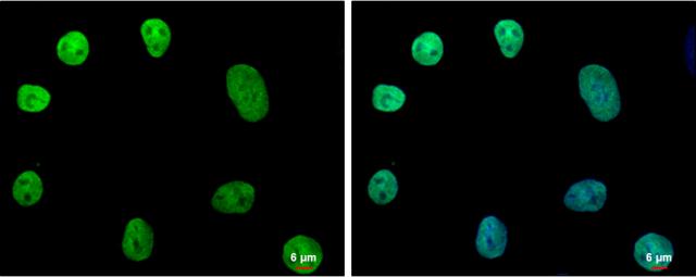
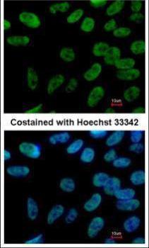
NANOG antibody [N3C3] detects NANOG protein at nucleus by immunofluorescent analysis. Sample: NT2D1 cells were fixed in 2% paraformaldehyde/culture medium at RT for 30 min. Green: NANOG protein stained by NANOG antibody [N3C3] diluted at 1:500. Blue: Hoechst 33342 staining. Scale bar = 6 μm.Application
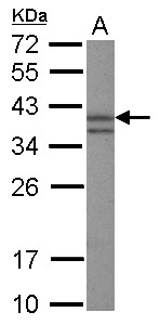
Sample (30 µg of whole cell lysate) A: HeLa nucleus 12% SDS PAGE diluted at 1:3000 The HRP-conjugated anti-rabbit IgG antibody was used to detect the primary antibody.Application
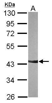
Sample (30 µg of whole cell lysate) A: NIH-3T3 10% SDS PAGE diluted at 1:3000 The HRP-conjugated anti-rabbit IgG antibody was used to detect the primary antibody.Application
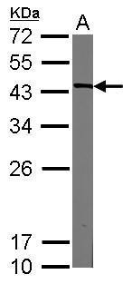
Sample (20 µg of whole cell lysate) A: mouse ESC 12% SDS PAGE diluted at 1:3000 The HRP-conjugated anti-rabbit IgG antibody was used to detect the primary antibody.Application
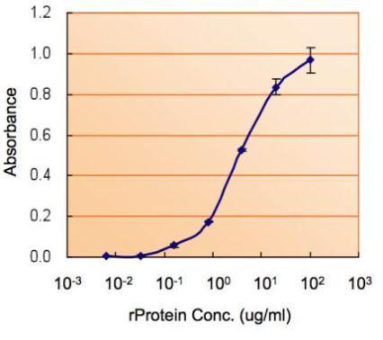
ELISA detection of NANOG using for capture at a concentration of 5 µg/mL and for detection at a concentration of 5 µg/mL.Application
Immunofluorescence analysis of paraformaldehyde-fixed Human ESC, using NANOG antibody at 1:400 dilution.Application
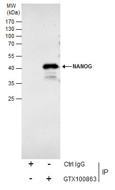
Immunoprecipitation of NANOG protein from NT2D1 whole cell extracts using 5 μg of NANOG antibody [N3C3] . Western blot analysis was performed using NANOG antibody [N3C3] . EasyBlot anti-Rabbit IgG was used as a secondary reagent.| Product Name | Nanog antibody |
|---|---|
| Antibody Type | Primary Antibodies |
| Product description | Rabbit Polyclonal antibody to Nanog (Nanog homeobox)Transcription regulator involved in inner cell mass and embryonic stem (ES) cells proliferation and self-renewal. Imposes pluripotency on ES cells and prevents their differentiation towards extraembryonic endoderm and trophectoderm lineages. Blocks bone morphogenetic protein-induced mesoderm differentiation of ES cells by physically interacting with SMAD1 and interfering with the recruitment of coactivators to the active SMAD transcriptional complexes (By similarity). Acts as a transcriptional activator or repressor (By similarity). Binds optimally to the DNA consensus sequence 5'-TAAT[GT][GT]-3' or 5'-[CG][GA][CG]C[GC]ATTAN[GC]-3' (By similarity). When overexpressed, promotes cells to enter into S phase and proliferation. |
| Immunogen | Recombinant protein encompassing a sequence within the center region of human NANOG. The exact sequence is proprietary. |
| Clonality | Polyclonal |
|---|---|
| Isotype | IgG |
| Host Species | Rabbit |
| Tested Applications | ELISAICC/IFIHCIHC-FIPWB |
| WB:1:200~1:1000 IP:1:50~1:250 ICC/IF:1:50~1:250 IHC:1:20~1:50 *Optimal dilutions/concentrations should be determined by the researcher.: |
|
| Species Reactivity | HumanMouse |
| Predicted Reactivity | ChimpanzeeChimpanzee (98%) |
| Concentration | 0.5mg/ml |
| Purification | Affinity purified |
| Gene Synonyms | NANOG antibody homeobox transcription factor Nanog-delta 48 antibody hNanog antibody homeobox protein NANOG antibody homeobox transcription factor Nanog antibody Nanog homeobox antibody |
|---|---|
| Molecular Weight(MW) | 35 kDa (note) (2) |
| Cellular Localization | Nucleus |
Application
NANOG antibody [N3C3] detects NANOG protein at nucleus by immunofluorescent analysis. Sample: NT2D1 cells were fixed in 2% paraformaldehyde/culture medium at RT for 30 min. Green: NANOG protein stained by NANOG antibody [N3C3] diluted at 1:500. Blue: Hoechst 33342 staining. Scale bar = 6 μm.
Application
Sample (30 µg of whole cell lysate) A: HeLa nucleus 12% SDS PAGE diluted at 1:3000 The HRP-conjugated anti-rabbit IgG antibody was used to detect the primary antibody.
Application
Sample (30 µg of whole cell lysate) A: NIH-3T3 10% SDS PAGE diluted at 1:3000 The HRP-conjugated anti-rabbit IgG antibody was used to detect the primary antibody.
Application
Sample (20 µg of whole cell lysate) A: mouse ESC 12% SDS PAGE diluted at 1:3000 The HRP-conjugated anti-rabbit IgG antibody was used to detect the primary antibody.
Application
ELISA detection of NANOG using for capture at a concentration of 5 µg/mL and for detection at a concentration of 5 µg/mL.
Application
Immunofluorescence analysis of paraformaldehyde-fixed Human ESC, using NANOG antibody at 1:400 dilution.
Application
Immunoprecipitation of NANOG protein from NT2D1 whole cell extracts using 5 μg of NANOG antibody [N3C3] . Western blot analysis was performed using NANOG antibody [N3C3] . EasyBlot anti-Rabbit IgG was used as a secondary reagent.| Positive Control | HeLa nucleus , NIH-3T3 , mouse ESC |
|---|---|
| Application Notes | WB:1:200~1:1000 IP:1:50~1:250 ICC/IF:1:50~1:250 IHC:1:20~1:50 *Optimal dilutions/concentrations should be determined by the researcher.: |
| Form | Liquid |
|---|---|
| Storage Instructions | Keep as concentrated solution. Aliquot and store at -20ºC or below. Avoid multiple freeze-thaw cycles. |
| Storage Buffer | 1XPBS, 1% BSA, 20% Glycerol (pH7). 0.01% Thimerosal was added as a preservative. |


