Application
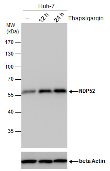
NDP52 antibody detects NDP52 protein by western blot analysis. Un-treated (-) and treated (+, Thapsigargin treatment for 12hrs and 24hrs) Huh-7 whole cell extracts (30 μg) were separated by 10% SDS-PAGE, and the membrane was blotted with NDP52 antibody diluted by 1:500. The ACTB was used as internal control ( 1:50000) shown at the bottom panel.Application
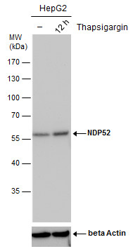
NDP52 antibody detects NDP52 protein by western blot analysis. Un-treated (-) and treated (+, Thapsigargin treatment for 12hrs) HepG2 whole cell extracts (30 μg) were separated by 10% SDS-PAGE, and the membrane was blotted with NDP52 antibody diluted by 1:500. The ACTB was used as internal control ( 1:50000) shown at the bottom panel.Application
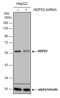
Non-transfected (–) and transfected (+) HepG2 whole cell extracts (30 µg) were separated by 10% SDS-PAGE, and the membrane was blotted with NDP52 antibody [GT422] diluted at 1:1000.Application
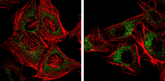
NDP52 antibody [GT422] detects NDP52 protein at autophagosome by immunofluorescent analysis. Samples: HeLa cells mock (left) and treated with 50μM Chloroquine for 24 hr (right) were fixed in 4% paraformaldehyde at RT for 15 min. Green: NDP52 protein stained by NDP52 antibody [GT422] diluted at 1:1000. Red: Phalloidin, a F-actin marker.Application
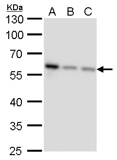
NDP52 antibody [GT422] detects NDP52 protein by western blot analysis. A. 30 μg Jurkat whole cell lysate/extract B. 30 μg Raji whole cell lysate/extract C. 30 μg NCI-H929 whole cell lysate/extract 10 % SDS-PAGE NDP52 antibody [GT422] dilution: 1:1000Application
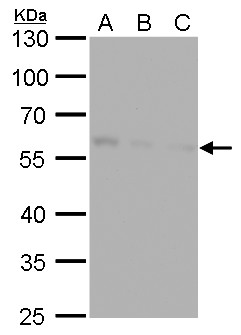
NDP52 antibody [GT422] detects NDP52 protein by western blot analysis. A. 30 μg Huh7 whole cell lysate/extract B. 30 μg Hep3B whole cell lysate/extract C. 30 μg HepG2 whole cell lysate/extract 10 % SDS-PAGE NDP52 antibody [GT422] dilution: 1:1000Application
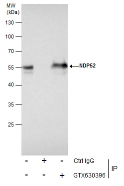
Immunoprecipitation of NDP52 protein from Jurkat whole cell extracts using 5 μg of NDP52 antibody [GT422] . Western blot analysis was performed using NDP52 antibody [GT422] . EasyBlot anti-Mouse IgG was used as a secondary reagent.Application
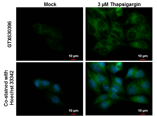
NDP52 antibody [GT422] detects NDP52 protein at autophagosome by immunofluorescent analysis. Samples: Hep G2 cells mock (left) and treated with 3 μM Thapsigargin for 16 hrs (right) were fixed in ice-cold MeOH for 10 min and permeabilized with 100% MeOH for 30 sec. Green: NDP52 protein stained by NDP52 antibody [GT422] diluted at 1:1000. Blue: Hoechst 33342 staining. Scale bar = 10 μm.| Product Name | NDP52 antibody |
|---|---|
| Antibody Type | Primary Antibodies |
| Product description | Mouse Monoclonal antibody [GT422] to NDP52 (calcium binding and coiled-coil domain 2)The protein encoded by this gene is a subunit of nuclear domain 10 (ND10) bodies. ND10 bodies are nuclear domains appearing immunohistochemically as ten dots per nucleus. They are believed to be associated with the nuclear matrix on the basis of their resistance to nuclease digestion and salt extraction. ND10 proteins are removed from the nucleus by herpes simplex virus-1 infection and may have a role in viral life cycles. [provided by RefSeq] |
| Immunogen | Recombinant protein encompassing a sequence within the center region of human NDP52. The exact sequence is proprietary. |
| Clonality | Monoclonal |
|---|---|
| Isotype | IgG1 |
| Clone Number | GT422 |
| Host Species | Mouse |
| Tested Applications | ICC/IFIPWB |
| *Optimal dilutions/concentrations should be determined by the researcher.
: |
|
| Species Reactivity | Human |
| Predicted Reactivity | ChimpanzeeChimpanzee (97%) |
| Concentration | 1mg/ml (Please refer to the vial label for the specific concentration) |
| Purification | Affinity purified |
| Gene Synonyms | calcium binding and coiled-coil domain 2 CALCOCO2 |
|---|---|
| Target | NDP52 |
| Cellular Localization | Cytoplasm , perinuclear region , Golgi apparatus , cytoskeleton |
Application
NDP52 antibody detects NDP52 protein by western blot analysis. Un-treated (-) and treated (+, Thapsigargin treatment for 12hrs and 24hrs) Huh-7 whole cell extracts (30 μg) were separated by 10% SDS-PAGE, and the membrane was blotted with NDP52 antibody diluted by 1:500. The ACTB was used as internal control ( 1:50000) shown at the bottom panel.
Application
NDP52 antibody detects NDP52 protein by western blot analysis. Un-treated (-) and treated (+, Thapsigargin treatment for 12hrs) HepG2 whole cell extracts (30 μg) were separated by 10% SDS-PAGE, and the membrane was blotted with NDP52 antibody diluted by 1:500. The ACTB was used as internal control ( 1:50000) shown at the bottom panel.
Application
Non-transfected (–) and transfected (+) HepG2 whole cell extracts (30 µg) were separated by 10% SDS-PAGE, and the membrane was blotted with NDP52 antibody [GT422] diluted at 1:1000.
Application
NDP52 antibody [GT422] detects NDP52 protein at autophagosome by immunofluorescent analysis. Samples: HeLa cells mock (left) and treated with 50μM Chloroquine for 24 hr (right) were fixed in 4% paraformaldehyde at RT for 15 min. Green: NDP52 protein stained by NDP52 antibody [GT422] diluted at 1:1000. Red: Phalloidin, a F-actin marker.
Application
NDP52 antibody [GT422] detects NDP52 protein by western blot analysis. A. 30 μg Jurkat whole cell lysate/extract B. 30 μg Raji whole cell lysate/extract C. 30 μg NCI-H929 whole cell lysate/extract 10 % SDS-PAGE NDP52 antibody [GT422] dilution: 1:1000
Application
NDP52 antibody [GT422] detects NDP52 protein by western blot analysis. A. 30 μg Huh7 whole cell lysate/extract B. 30 μg Hep3B whole cell lysate/extract C. 30 μg HepG2 whole cell lysate/extract 10 % SDS-PAGE NDP52 antibody [GT422] dilution: 1:1000
Application
Immunoprecipitation of NDP52 protein from Jurkat whole cell extracts using 5 μg of NDP52 antibody [GT422] . Western blot analysis was performed using NDP52 antibody [GT422] . EasyBlot anti-Mouse IgG was used as a secondary reagent.
Application
NDP52 antibody [GT422] detects NDP52 protein at autophagosome by immunofluorescent analysis. Samples: Hep G2 cells mock (left) and treated with 3 μM Thapsigargin for 16 hrs (right) were fixed in ice-cold MeOH for 10 min and permeabilized with 100% MeOH for 30 sec. Green: NDP52 protein stained by NDP52 antibody [GT422] diluted at 1:1000. Blue: Hoechst 33342 staining. Scale bar = 10 μm.| Positive Control | Jurkat , Raji , NCI-H929 , Huh-7 , Hep3B , HepG2 , Huh-7 (untreated) , Huh-7 (3 µM Thapsigargin treatment for 12 hr) , Huh-7 (3 µM Thapsigargin treatment for 24 hr) , HepG2 (untreated) , HepG2 (3 µM Thapsigargin treatment for 12 hr) |
|---|---|
| Application Notes | *Optimal dilutions/concentrations should be determined by the researcher.
: |
| Form | Liquid |
|---|---|
| Storage Instructions | Keep as concentrated solution. Aliquot and store at -20ºC or below. Avoid multiple freeze-thaw cycles. |
| Storage Buffer | PBS |


